1MDM | pdb_00001mdm

INHIBITED FRAGMENT OF ETS-1 AND PAIRED DOMAIN OF PAX5 BOUND TO DNA


Experimental Data Snapshot

  • Method: X-RAY DIFFRACTION
  • Resolution: 2.80 Å
  • R-Value Free: 
    0.310 (Depositor), 0.307 (DCC) 
  • R-Value Work: 
    0.259 (Depositor), 0.260 (DCC) 
  • R-Value Observed: 
    0.259 (Depositor) 

Starting Model: experimental
View more details

wwPDB Validation 3D Report Full Report

Validation slider image for 1MDM

This is version 1.3 of the entry. See complete history

Literature

STRUCTURAL ANALYSIS OF THE AUTOINHIBITION OF ETS-1 AND ITS ROLE IN PROTEIN PARTNERSHIPS

Garvie, C.W.Pufall, M.A.Graves, B.J.Wolberger, C.

(2002) J Biological Chem 277: 45529-45536

  • DOI: https://doi.org/10.1074/jbc.M206327200
  • Primary Citation Related Structures: 
    1MD0, 1MDM

  • PubMed Abstract: 

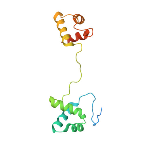
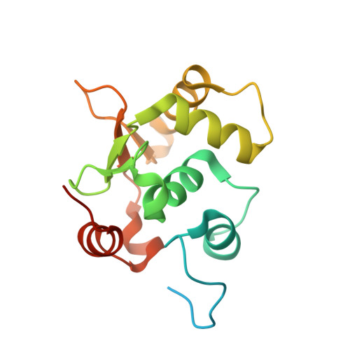
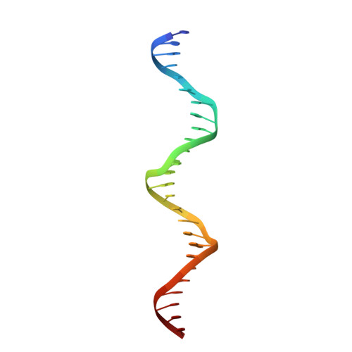
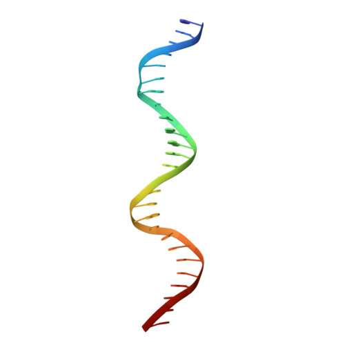
    The DNA-binding activity of the eukaryotic transcription factor Ets-1 (E26 avian erythroblastosis virus oncogene-E twenty-six) is negatively regulated by inhibitory regions that flank the ETS domain. Based on the results of solution studies, these N- and C-terminal inhibitory regions have been proposed to pack against the ETS domain and form an autoinhibitory module whose N terminus partially unfolds upon binding of Ets-1 to DNA. Mutations that disrupt autoinhibition of DNA binding also cause a structural change in the inhibitory region. We report here a crystallographic study of fragments of Ets-1 that provide structural details of the inhibitory module and the structural transition that accompanies DNA binding. The structures of free and DNA-bound Ets-1 fragments containing the ETS domain and the inhibitory regions confirm that the N-terminal inhibitory region contains two alpha-helices one of which unfolds upon Ets-1 binding to DNA. The observations from the crystal structure, coupled with mutagenesis experiments, allow us to propose a model for the inhibited form of Ets-1 and lend insight into the flexible interaction between Ets-1 and the acute myeloid leukemia 1 protein, AML1 (RUNX1).


  • Organizational Affiliation
    • Department of Biophysics and Biophysical Chemistry and the Howard Hughes Medical Institute, Johns Hopkins University School of Medicine, Baltimore, Maryland 21205-2185, USA.

Macromolecule Content 

  • Total Structure Weight: 51.31 kDa 
  • Atom Count: 3,054 
  • Modeled Residue Count: 305 
  • Deposited Residue Count: 362 
  • Unique protein chains: 2
  • Unique nucleic acid chains: 2

Macromolecules


Find similar proteins by:|  3D Structure
Entity ID: 3
MoleculeChains  Sequence LengthOrganismDetailsImage
PAIRED BOX PROTEIN PAX-5C [auth A]149Homo sapiensMutation(s): 0 
Gene Names: Pax5
UniProt & NIH Common Fund Data Resources
Find proteins for Q02548 (Homo sapiens)
Explore Q02548 
Go to UniProtKB:  Q02548
PHAROS:  Q02548
GTEx:  ENSG00000196092 
Entity Groups
Sequence Clusters30% Identity50% Identity70% Identity90% Identity95% Identity100% Identity
UniProt GroupQ02548
Sequence Annotations
Expand
Reference Sequence
Find similar proteins by:|  3D Structure
Entity ID: 4
MoleculeChains  Sequence LengthOrganismDetailsImage
C-ETS-1 PROTEIND [auth B]161Mus musculusMutation(s): 0 
Gene Names: Ets-1
UniProt
Find proteins for P27577 (Mus musculus)
Explore P27577 
Go to UniProtKB:  P27577
Entity Groups
Sequence Clusters30% Identity50% Identity70% Identity90% Identity95% Identity100% Identity
UniProt GroupP27577
Sequence Annotations
Expand
Reference Sequence
Find similar nucleic acids by:  (by identity cutoff) 
Entity ID: 1
MoleculeChains LengthOrganismImage
PAX5/ETS BINDING SITE ON THE MB-1 PROMOTERA [auth C]26N/A
Sequence Annotations
Expand
Reference Sequence
Find similar nucleic acids by:  (by identity cutoff) 
Entity ID: 2
MoleculeChains LengthOrganismImage
PAX5/ETS BINDING SITE ON THE MB-1 PROMOTERB [auth D]26N/A
Sequence Annotations
Expand
Reference Sequence

Experimental Data & Validation

Experimental Data

  • Method: X-RAY DIFFRACTION
  • Resolution: 2.80 Å
  • R-Value Free:  0.310 (Depositor), 0.307 (DCC) 
  • R-Value Work:  0.259 (Depositor), 0.260 (DCC) 
  • R-Value Observed: 0.259 (Depositor) 
Space Group: P 21 21 2
Unit Cell:
Length ( Å )Angle ( ˚ )
a = 77.27α = 90
b = 171.23β = 90
c = 44.71γ = 90
Software Package:
Software NamePurpose
MOSFLMdata reduction
SCALAdata scaling
AMoREphasing
CNSrefinement
CCP4data scaling

Structure Validation

View Full Validation Report


Entry History 

Deposition Data

Revision History  (Full details and data files)

  • Version 1.0: 2002-12-11
    Type: Initial release
  • Version 1.1: 2008-04-28
    Changes: Version format compliance
  • Version 1.2: 2011-07-13
    Changes: Version format compliance
  • Version 1.3: 2024-02-14
    Changes: Data collection, Database references, Refinement description