1MDA

CRYSTAL STRUCTURE OF AN ELECTRON-TRANSFER COMPLEX BETWEEN METHYLAMINE DEHYDROGENASE AND AMICYANIN


Experimental Data Snapshot

  • Method: X-RAY DIFFRACTION
  • Resolution: 2.50 Å
  • R-Value Observed: 0.285 

wwPDB Validation   3D Report Full Report


This is version 1.2 of the entry. See complete history


Literature

Crystal structure of an electron-transfer complex between methylamine dehydrogenase and amicyanin.

Chen, L.Durley, R.Poliks, B.J.Hamada, K.Chen, Z.Mathews, F.S.Davidson, V.L.Satow, Y.Huizinga, E.Vellieux, F.M.Hol, W.G.J.

(1992) Biochemistry 31: 4959-4964

  • DOI: https://doi.org/10.1021/bi00136a006
  • Primary Citation of Related Structures:  
    1MDA

  • PubMed Abstract: 

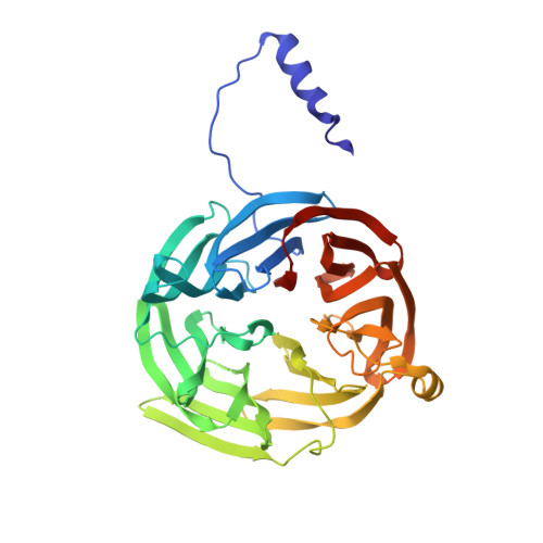
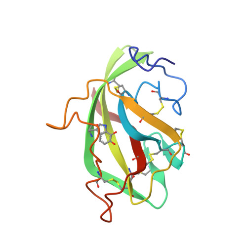
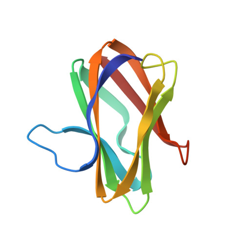
    The crystal structure of the complex between the quinoprotein methylamine dehydrogenase (MADH) and the type I blue copper protein amicyanin, both from Paracoccus denitrificans, has been determined at 2.5-A resolution using molecular replacement. The search model was MADH from Thiobacillus versutus. The amicyanin could be located in an averaged electron density difference map and the model improved by refinement and model building procedures. Nine beta-strands are observed within the amicyanin molecule. The copper atom is located between three antiparallel strands and is about 2.5 A below the protein surface. The major intermolecular interactions occur between amicyanin and the light subunit of MADH where the interface is largely hydrophobic. The copper atom of amicyanin and the redox cofactor of MADH are about 9.4 A apart. One of the copper ligands, His 95, lies between the two redox centers and may facilitate electron transfer between them.


  • Organizational Affiliation

    Department of Cell Biology and Physiology, Washington University School of Medicine, St. Louis, Missouri 63110.


Macromolecules
Find similar proteins by:  (by identity cutoff)  |  3D Structure
Entity ID: 1
MoleculeChains Sequence LengthOrganismDetailsImage
METHYLAMINE DEHYDROGENASE (HEAVY SUBUNIT)A [auth H],
C [auth J]
368Paracoccus denitrificansMutation(s): 0 
EC: 1.4.99.3
Entity Groups  
Sequence Clusters30% Identity50% Identity70% Identity90% Identity95% Identity100% Identity
Sequence Annotations
Expand
  • Reference Sequence
Find similar proteins by:  (by identity cutoff)  |  3D Structure
Entity ID: 2
MoleculeChains Sequence LengthOrganismDetailsImage
METHYLAMINE DEHYDROGENASE (LIGHT SUBUNIT)B [auth L],
D [auth M]
121Paracoccus denitrificansMutation(s): 0 
EC: 1.4.99.3
Entity Groups  
Sequence Clusters30% Identity50% Identity70% Identity90% Identity95% Identity100% Identity
Sequence Annotations
Expand
  • Reference Sequence
Find similar proteins by:  (by identity cutoff)  |  3D Structure
Entity ID: 3
MoleculeChains Sequence LengthOrganismDetailsImage
AMICYANINE [auth A],
F [auth B]
103Paracoccus denitrificansMutation(s): 0 
UniProt
Find proteins for P22364 (Paracoccus denitrificans)
Explore P22364 
Go to UniProtKB:  P22364
Entity Groups  
Sequence Clusters30% Identity50% Identity70% Identity90% Identity95% Identity100% Identity
UniProt GroupP22364
Sequence Annotations
Expand
  • Reference Sequence
Small Molecules
Ligands 1 Unique
IDChains Name / Formula / InChI Key2D Diagram3D Interactions
CU
Query on CU

Download Ideal Coordinates CCD File 
G [auth A],
H [auth B]
COPPER (II) ION
Cu
JPVYNHNXODAKFH-UHFFFAOYSA-N
Modified Residues  1 Unique
IDChains TypeFormula2D DiagramParent
TRQ
Query on TRQ
B [auth L],
D [auth M]
L-PEPTIDE LINKINGC11 H10 N2 O4TRP
Experimental Data & Validation

Experimental Data

  • Method: X-RAY DIFFRACTION
  • Resolution: 2.50 Å
  • R-Value Observed: 0.285 
  • Space Group: P 41 21 2
Unit Cell:
Length ( Å )Angle ( ˚ )
a = 124.7α = 90
b = 124.7β = 90
c = 247.4γ = 90
Software Package:
Software NamePurpose
PROLSQrefinement
TNTrefinement

Structure Validation

View Full Validation Report


Entry History 

Deposition Data

Revision History  (Full details and data files)

  • Version 1.0: 1993-10-31
    Type: Initial release
  • Version 1.1: 2008-03-03
    Changes: Version format compliance
  • Version 1.2: 2011-07-13
    Changes: Version format compliance