Comparison of the free and DNA-complexed forms of the DNA-binding domain from c-Myb.
Ogata, K., Morikawa, S., Nakamura, H., Hojo, H., Yoshimura, S., Zhang, R., Aimoto, S., Ametani, Y., Hirata, Z., Sarai, A., Ishii, S., Nishimura, Y.(1995) Nat Struct Biol 2: 309-320
- PubMed: 7796266 Search on PubMed
- DOI: https://doi.org/10.1038/nsb0495-309
- Primary Citation Related Structures:
1MBE, 1MBF, 1MBG, 1MBH, 1MBJ, 1MBK - PubMed Abstract:
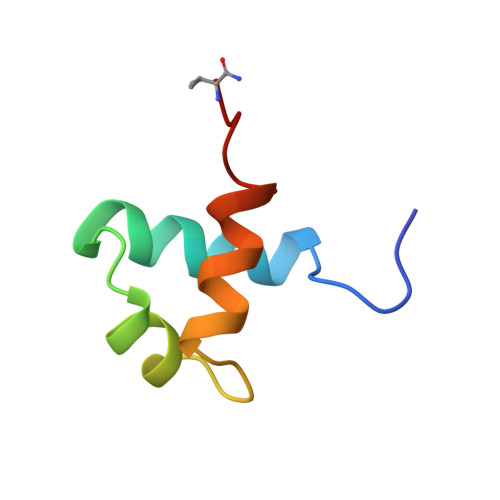
The DNA-binding domain of c-Myb consists of three imperfect tandem repeats (R1, R2 and R3). The three repeats have similar overall architectures, each containing a helix-turn-helix variation motif. The three conserved tryptophans in each repeat participate in forming a hydrophobic core. Comparison of the three repeat structures indicated that cavities are found in the hydrophobic core of R2, which is thermally unstable. On complexation with DNA, the orientations of R2 and R3 are fixed by tight binding and their conformations are slightly changed. No significant changes occur in the chemical shifts of R1 consistent with its loose interaction with DNA.
- Graduate School of Integrated Science, Yokohama City University, Japan.
Organizational Affiliation: