1LQO

Crystal Strutcure of the Fosfomycin Resistance Protein A (FosA) Containing Bound Thallium Cations


Experimental Data Snapshot

  • Method: X-RAY DIFFRACTION
  • Resolution: 2.00 Å
  • R-Value Free: 0.252 
  • R-Value Work: 0.197 
  • R-Value Observed: 0.200 

wwPDB Validation   3D Report Full Report


This is version 1.3 of the entry. See complete history


Literature

Crystal structure of a genomically encoded fosfomycin resistance protein (FosA) at 1.19 A resolution by MAD phasing off the L-III edge of Tl(+)

Rife, C.L.Pharris, R.E.Newcomer, M.E.Armstrong, R.N.

(2002) J Am Chem Soc 124: 11001-11003

  • DOI: https://doi.org/10.1021/ja026879v
  • Primary Citation of Related Structures:  
    1LQK, 1LQO, 1LQP

  • PubMed Abstract: 

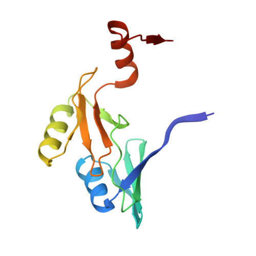
    The fosfomycin resistance protein (FosA) catalyzes the Mn(II)- and K+-dependent addition of glutathione to the oxirane of the antibiotic fosfomycin. The crystal structure of FosA from Pseudomonas aeruginosa was solved at a resolution of 1.19 A by multiwavelength anomalous diffraction at the L-III edge of a Tl+ derivative. The structure solution took advantage of the ability of Tl+ to substitute for K+. The existence of multiple Tl sites in the asymmetric unit suggests that this may be a generally useful technique for phasing protein crystal structures. A 1.35 A resolution structure with phosphate bound in the active site shows that the Mn(II) center has a rare four-coordinate geometry. The structure of the fosfomycin complex at 1.19 A resolution indicates that the Mn(II) center is close to five-coordinate with trigonal bipyramidal geometry and a ligand set consisting of two histidines (H7 and H64) and one phosphonate oxygen occupying the equatorial sites and the carboxylate of E110 at one of the apical sites. The oxirane oxygen of the substrate is located at the other apical site but is 0.2 A beyond the average Mn-O distance for five-coordinate Mn(II). The Mn(II) center is proposed to stabilize the alkoxide in the transition state, while the nearby hydroxyl group of T9 acts as a proton donor in the reaction. The K+ ion located 6.5 A from the Mn(II) appears to help orient the substrate for nucleophilic attack.


  • Organizational Affiliation

    Departments of Biochemistry and Chemistry, Vanderbilt University, Nashville, Tennessee 37232-0146, USA.


Macromolecules
Find similar proteins by:  (by identity cutoff)  |  3D Structure
Entity ID: 1
MoleculeChains Sequence LengthOrganismDetailsImage
PROBABLE Fosfomycin Resistance Protein
A, B
135Pseudomonas aeruginosaMutation(s): 0 
Gene Names: FosA
EC: 2.5.1.18
UniProt
Find proteins for Q9I4K6 (Pseudomonas aeruginosa (strain ATCC 15692 / DSM 22644 / CIP 104116 / JCM 14847 / LMG 12228 / 1C / PRS 101 / PAO1))
Explore Q9I4K6 
Go to UniProtKB:  Q9I4K6
Entity Groups  
Sequence Clusters30% Identity50% Identity70% Identity90% Identity95% Identity100% Identity
UniProt GroupQ9I4K6
Sequence Annotations
Expand
  • Reference Sequence
Small Molecules
Ligands 3 Unique
IDChains Name / Formula / InChI Key2D Diagram3D Interactions
TL
Query on TL

Download Ideal Coordinates CCD File 
G [auth A]
H [auth A]
I [auth A]
L [auth B]
M [auth B]
G [auth A],
H [auth A],
I [auth A],
L [auth B],
M [auth B],
N [auth B]
THALLIUM (I) ION
Tl
ZLUSCZLCHQSJRU-UHFFFAOYSA-N
PO4
Query on PO4

Download Ideal Coordinates CCD File 
C [auth A],
D [auth A],
E [auth A],
J [auth B]
PHOSPHATE ION
O4 P
NBIIXXVUZAFLBC-UHFFFAOYSA-K
MN
Query on MN

Download Ideal Coordinates CCD File 
F [auth A],
K [auth B]
MANGANESE (II) ION
Mn
WAEMQWOKJMHJLA-UHFFFAOYSA-N
Experimental Data & Validation

Experimental Data

  • Method: X-RAY DIFFRACTION
  • Resolution: 2.00 Å
  • R-Value Free: 0.252 
  • R-Value Work: 0.197 
  • R-Value Observed: 0.200 
  • Space Group: P 21 21 21
Unit Cell:
Length ( Å )Angle ( ˚ )
a = 55.797α = 90
b = 65.874β = 90
c = 76.958γ = 90
Software Package:
Software NamePurpose
DENZOdata reduction
SCALEPACKdata scaling
SHARPphasing
REFMACrefinement

Structure Validation

View Full Validation Report


Entry History 

Deposition Data

Revision History  (Full details and data files)

  • Version 1.0: 2002-09-11
    Type: Initial release
  • Version 1.1: 2008-04-28
    Changes: Version format compliance
  • Version 1.2: 2011-07-13
    Changes: Version format compliance
  • Version 1.3: 2024-02-14
    Changes: Advisory, Data collection, Database references, Derived calculations