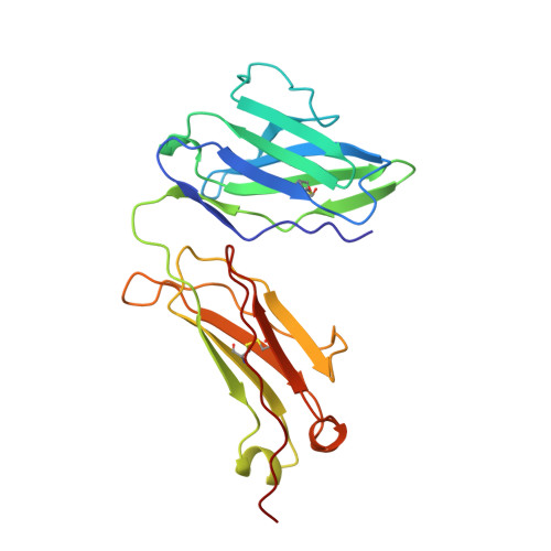
Pitfalls of molecular replacement: the structure determination of an immunoglobulin light-chain dimer.
Huang, D.B., Ainsworth, C., Solomon, A., Schiffer, M.(1996) Acta Crystallogr D Biol Crystallogr 52: 1058-1066
- PubMed: 15299564
- DOI: https://doi.org/10.1107/S090744499600813X
- Primary Citation of Related Structures:
1LIL - PubMed Abstract:
The structure of protein Cle, a human light-chain dimer from the lambdaIII subgroup, was determined using 2.6 A data; the R value is 18.4%. The structure was solved, after a false start, by molecular replacement with the lambdaII/V Mcg protein as a search structure. When the refinement did not proceed beyond an R value of 27%, it was discovered that while the constant domains were in their correct positions in the unit cell, the incorrect variable domains were used for defining the molecule. The correct solution required a rotation of 180 degrees around the local twofold axis that relates the two constant domains of the dimer. The correct variable domain positions overlap about 70% of the same volume as the incorrect ones of a symmetry-related molecule. The refinement distorted the geometries of the domains. Though the constant domains were in their correct positions, the r.m.s. (root-mean-square) deviation of the Calpha atom position was 1.2 A when the two constant domains were compared. For the correct structure, this value is 0.5 A. The phi and psi angles, the r.m.s. chiral value and the free R value, even when calculated a posteriori, were good indicators of the correctness of the structure. The quaternary structure of the Cle molecule is similar to that in Mcg (crystallized from ammonium sulfate); the elbow bend is 115 degrees. However, the arrangement of the variable domains differs from that observed in other variable domain dimers. The variable domains of Cle are 0.7 A closer than in Mcg or variable dimer Rei. The hydrogen bonding at the interface of the two domains is novel. Residues Tyr36 from both monomers form a hydrogen bond that is part of a network with the Gln89 residues from both monomers. For the first time hydrogen bonds were observed between the main-chain peptide N and O atoms of the complementarity-determining region CDR2 and CDR3 segments of both monomers.
- Center for Mechanistic Biology and Biotechnology, Argonne National Laboratory, Illinois 60439-4833, USA.
Organizational Affiliation:


