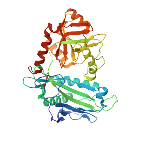
Crystal structures of human mitochondrial branched chain aminotransferase reaction intermediates: ketimine and pyridoxamine phosphate forms
Yennawar, N.H., Conway, M.E., Yennawar, H.P., Farber, G.K., Hutson, S.M.(2002) Biochemistry 41: 11592-11601
- PubMed: 12269802 Search on PubMed
- DOI: https://doi.org/10.1021/bi020221c
- Primary Citation Related Structures:
1KT8, 1KTA - PubMed Abstract:
The three-dimensional structures of the isoleucine ketimine and the pyridoxamine phosphate forms of human mitochondrial branched chain aminotransferase (hBCATm) have been determined crystallographically at 1.9 A resolution. The hBCATm-catalyzed transamination can be described in molecular terms together with the earlier solved pyridoxal phosphate forms of the enzyme. The active site lysine, Lys202, undergoes large conformational changes, and the pyridine ring of the cofactor tilts by about 18 degrees during catalysis. A major determinant of the enzyme's substrate and stereospecificity for L-branched chain amino acids is a group of hydrophobic residues that form three hydrophobic surfaces and lock the side chain in place. Short-chain aliphatic amino acid side chains are unable to interact through van der Waals contacts with any of the surfaces whereas bulky aromatic side chains would result in significant steric hindrance. As shown by modeling, and in agreement with previous biochemical data, glutamate but not aspartate can form hydrogen bond interactions. The carboxylate group of the bound isoleucine is on the same side as the phosphate group of the cofactor. These active site interactions are largely retained in a model of the human cytosolic branched chain aminotransferase (hBCATc), suggesting that residues in the second tier of interactions are likely to determine the specificity of hBCATc for the drug gabapentin. Finally, the structures reveal a unique role for cysteine residues in the mammalian BCAT. Cys315 and Cys318, which immediately follow a beta-turn (residues 311-314) and are located just outside the active site, form an unusual thiol-thiolate hydrogen bond. This beta-turn positions Thr313 for its interaction with the pyridoxal phosphate oxygens and substrate alpha-carboxylate group.
- Department of Biochemistry and Molecular Biology, Althouse Laboratory, The Pennsylvania State University, University Park, PA 16802, USA. nhy1@psu.edu
Organizational Affiliation: