1KQR

Crystal Structure of the Rhesus Rotavirus VP4 Sialic Acid Binding Domain in Complex with 2-O-methyl-alpha-D-N-acetyl neuraminic acid


Experimental Data Snapshot

  • Method: X-RAY DIFFRACTION
  • Resolution: 1.40 Å
  • R-Value Free: 0.181 
  • R-Value Work: 0.169 

wwPDB Validation   3D Report Full Report


Ligand Structure Quality Assessment 


This is version 1.3 of the entry. See complete history


Literature

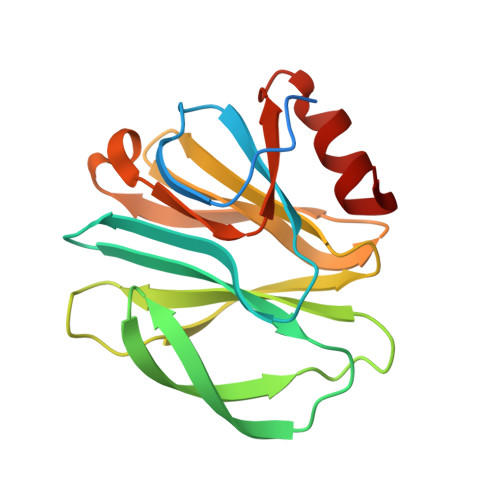
The Rhesus Rotavirus VP4 Sialic Acid Binding Domain has a Galectin Fold with a Novel Carbohydrate Binding Site

Dormitzer, P.R.Sun, Z.-Y.J.Wagner, G.Harrison, S.C.

(2002) EMBO J 21: 885-897

  • DOI: https://doi.org/10.1093/emboj/21.5.885
  • Primary Citation of Related Structures:  
    1KQR, 1KRI

  • PubMed Abstract: 
  • Cell attachment and membrane penetration are functions of the rotavirus outer capsid spike protein, VP4. An activating tryptic cleavage of VP4 produces the N-terminal fragment, VP8*, which is the viral hemagglutinin and an important target of neutralizing antibodies ...

    Cell attachment and membrane penetration are functions of the rotavirus outer capsid spike protein, VP4. An activating tryptic cleavage of VP4 produces the N-terminal fragment, VP8*, which is the viral hemagglutinin and an important target of neutralizing antibodies. We have determined, by X-ray crystallography, the atomic structure of the VP8* core bound to sialic acid and, by NMR spectroscopy, the structure of the unliganded VP8* core. The domain has the beta-sandwich fold of the galectins, a family of sugar binding proteins. The surface corresponding to the galectin carbohydrate binding site is blocked, and rotavirus VP8* instead binds sialic acid in a shallow groove between its two beta-sheets. There appears to be a small induced fit on binding. The residues that contact sialic acid are conserved in sialic acid-dependent rotavirus strains. Neutralization escape mutations are widely distributed over the VP8* surface and cluster in four epitopes. From the fit of the VP8* core into the virion spikes, we propose that VP4 arose from the insertion of a host carbohydrate binding domain into a viral membrane interaction protein.


    Related Citations: 
    • Proteolysis of Monomeric Recombinant Rotavirus VP4 Yields an Oligomeric VP5* Core
      Dormitzer, P.R., Greenberg, H.B., Harrison, S.C.
      (2001) J Virol 75: 7339

    Organizational Affiliation

    Laboratory of Molecular Medicine, Enders 673, Children's Hospital, 320 Longwood Avenue, Boston, MA 02115, USA. dormitze@crystal.harvard.edu


Macromolecules
Find similar proteins by:  (by identity cutoff)  |  3D Structure
Entity ID: 1
MoleculeChainsSequence LengthOrganismDetailsImage
VP4179Simian rotavirus A strain RRVMutation(s): 0 
Gene Names: segment 4
UniProt
Find proteins for P12473 (Rotavirus A (strain RVA/Monkey/United States/RRV/1975/G3P5B[3]))
Explore P12473 
Go to UniProtKB:  P12473
Entity Groups  
Sequence Clusters30% Identity50% Identity70% Identity90% Identity95% Identity100% Identity
UniProt GroupP12473
Protein Feature View
Expand
  • Reference Sequence
Small Molecules
Ligands 3 Unique
IDChainsName / Formula / InChI Key2D Diagram3D Interactions
MNA
Query on MNA

Download Ideal Coordinates CCD File 
C [auth A]2-O-methyl-5-N-acetyl-alpha-D-neuraminic acid
C12 H21 N O9
NJRVVFURCKKXOD-MIDKXNQYSA-N
 Ligand Interaction
SO4
Query on SO4

Download Ideal Coordinates CCD File 
B [auth A]SULFATE ION
O4 S
QAOWNCQODCNURD-UHFFFAOYSA-L
 Ligand Interaction
GOL
Query on GOL

Download Ideal Coordinates CCD File 
D [auth A]GLYCEROL
C3 H8 O3
PEDCQBHIVMGVHV-UHFFFAOYSA-N
 Ligand Interaction
Experimental Data & Validation

Experimental Data

  • Method: X-RAY DIFFRACTION
  • Resolution: 1.40 Å
  • R-Value Free: 0.181 
  • R-Value Work: 0.169 
  • Space Group: P 41 21 2
Unit Cell:
Length ( Å )Angle ( ˚ )
a = 48.027α = 90
b = 48.027β = 90
c = 130.404γ = 90
Software Package:
Software NamePurpose
CNSrefinement
DENZOdata reduction
HKL-2000data scaling
CNSphasing

Structure Validation

View Full Validation Report


Ligand Structure Quality Assessment 


Entry History 

Deposition Data

Revision History  (Full details and data files)

  • Version 1.0: 2002-03-27
    Type: Initial release
  • Version 1.1: 2008-04-27
    Changes: Version format compliance
  • Version 1.2: 2011-07-13
    Changes: Non-polymer description, Version format compliance
  • Version 1.3: 2020-07-29
    Type: Remediation
    Reason: Carbohydrate remediation
    Changes: Advisory, Data collection, Database references, Derived calculations, Structure summary