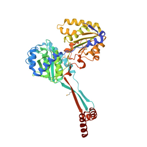
Mechanism of action and NAD+-binding mode revealed by the crystal structure of L-histidinol dehydrogenase.
Barbosa, J.A.R.G., Sivaraman, J., Li, Y., Larocque, R., Matte, A., Schrag, J.D., Cygler, M.(2002) Proc Natl Acad Sci U S A 99: 1859-1864
- PubMed: 11842181 Search on PubMedSearch on PubMed Central
- DOI: https://doi.org/10.1073/pnas.022476199
- Primary Citation Related Structures:
1K75, 1KAE, 1KAH, 1KAR - PubMed Abstract:
The histidine biosynthetic pathway is an ancient one found in bacteria, archaebacteria, fungi, and plants that converts 5-phosphoribosyl 1-pyrophosphate to l-histidine in 10 enzymatic reactions. This pathway provided a paradigm for the operon, transcriptional regulation of gene expression, and feedback inhibition of a pathway. l-histidinol dehydrogenase (HisD, EC ) catalyzes the last two steps in the biosynthesis of l-histidine: sequential NAD-dependent oxidations of l-histidinol to l-histidinaldehyde and then to l-histidine. HisD functions as a homodimer and requires the presence of one Zn(2+) cation per monomer. We have determined the three-dimensional structure of Escherichia coli HisD in the apo state as well as complexes with substrate, Zn(2+), and NAD(+) (best resolution is 1.7 A). Each monomer is made of four domains, whereas the intertwined dimer possibly results from domain swapping. Two domains display a very similar incomplete Rossmann fold that suggests an ancient event of gene duplication. Residues from both monomers form the active site. Zn(2+) plays a crucial role in substrate binding but is not directly involved in catalysis. The active site residue His-327 participates in acid-base catalysis, whereas Glu-326 activates a water molecule. NAD(+) binds weakly to one of the Rossmann fold domains in a manner different from that previously observed for other proteins having a Rossmann fold.
- Biotechnology Research Institute, National Research Council of Canada, 6100 Royalmount Avenue, Montreal, QC, Canada H4P 2R2.
Organizational Affiliation: