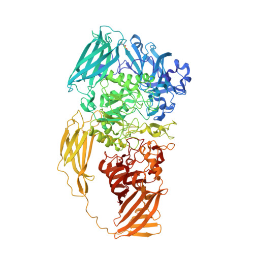
1JZ7

E. COLI (lacZ) BETA-GALACTOSIDASE IN COMPLEX WITH GALACTOSE


Experimental Data Snapshot

  • Method: X-RAY DIFFRACTION
  • Resolution: 1.50 Å
  • R-Value Free: 0.219 
  • R-Value Work: 0.176 

wwPDB Validation   3D Report Full Report


Ligand Structure Quality Assessment 


This is version 1.3 of the entry. See complete history


Literature

A Structural View of the Action of Escherichia Coli (Lacz) Beta-Galactosidase

Juers, D.H.Heightman, T.D.Vasella, A.McCarter, J.D.Mackenzie, L.Withers, S.G.Matthews, B.W.

(2001) Biochemistry 40: 14781-14794

  • DOI: https://doi.org/10.1021/bi011727i
  • Primary Citation of Related Structures:  
    1JYN, 1JYV, 1JYW, 1JYX, 1JZ2, 1JZ3, 1JZ4, 1JZ5, 1JZ6, 1JZ7, 1JZ8, 4V44, 4V45

  • PubMed Abstract: 
  • The structures of a series of complexes designed to mimic intermediates along the reaction coordinate for beta-galactosidase are presented. These complexes clarify and enhance previous proposals regarding the catalytic mechanism. The nucleophile, Glu537, is seen to covalently bind to the galactosyl moiety ...

    The structures of a series of complexes designed to mimic intermediates along the reaction coordinate for beta-galactosidase are presented. These complexes clarify and enhance previous proposals regarding the catalytic mechanism. The nucleophile, Glu537, is seen to covalently bind to the galactosyl moiety. Of the two potential acids, Mg(2+) and Glu461, the latter is in better position to directly assist in leaving group departure, suggesting that the metal ion acts in a secondary role. A sodium ion plays a part in substrate binding by directly ligating the galactosyl 6-hydroxyl. The proposed reaction coordinate involves the movement of the galactosyl moiety deep into the active site pocket. For those ligands that do bind deeply there is an associated conformational change in which residues within loop 794-804 move up to 10 A closer to the site of binding. In some cases this can be inhibited by the binding of additional ligands. The resulting restricted access to the intermediate helps to explain why allolactose, the natural inducer for the lac operon, is the preferred product of transglycosylation.


    Related Citations: 
    • High Resolution Structure of Beta-Galactosidase in a New Crystal Form Reveals Multiple Metal-Binding Sites and Provides a Structural Basis for Alpha-Complementation
      Juers, D.H., Jacobson, R.H., Wigley, D., Zhang, X.J., Huber, R.E., Tronrud, D.E., Matthews, B.W.
      (2000) Protein Sci 9: 1685
    • Structural Comparisons of Tim Barrel Proteins Suggest Functional and Evolutionary Relationships between Beta-Galactosidase and Other Glycohydrolases
      Juers, D.H., Huber, R.E., Matthews, B.W.
      (1999) Protein Sci 8: 122
    • Three-Dimensional Structure of Beta-Galactosidase from E. Coli
      Jacobson, R.H., Zhang, X.J., Dubose, R.F., Matthews, B.W.
      (1994) Nature 369: 761
    • Crystallization of beta-galactosidase from Escherichia coli
      Jacobson, R.H., Matthews, B.W.
      (1992) J Mol Biol 223: 1177

    Organizational Affiliation

    Institute of Molecular Biology, Howard Hughes Medical Institute and Department of Physics, University of Oregon, Eugene, Oregon 97403-1229, USA.


Macromolecules
Find similar proteins by:  (by identity cutoff)  |  3D Structure
Entity ID: 1
MoleculeChainsSequence LengthOrganismDetailsImage
Beta-GalactosidaseA, B, C, D1,023Escherichia coliMutation(s): 1 
Gene Names: lacZ
EC: 3.2.1.23
UniProt
Find proteins for P00722 (Escherichia coli (strain K12))
Explore P00722 
Go to UniProtKB:  P00722
Entity Groups  
Sequence Clusters30% Identity50% Identity70% Identity90% Identity95% Identity100% Identity
UniProt GroupP00722
Protein Feature View
Expand
  • Reference Sequence
Small Molecules
Ligands 4 Unique
IDChainsName / Formula / InChI Key2D Diagram3D Interactions
GAL
Query on GAL

Download Ideal Coordinates CCD File 
E [auth A],
F [auth A],
LA [auth B],
MA [auth B],
QB [auth C],
E [auth A],
F [auth A],
LA [auth B],
MA [auth B],
QB [auth C],
RB [auth C],
XC [auth D],
YC [auth D]
beta-D-galactopyranose
C6 H12 O6
WQZGKKKJIJFFOK-FPRJBGLDSA-N
 Ligand Interaction
DMS
Query on DMS

Download Ideal Coordinates CCD File 
AA [auth A],
AB [auth B],
AC [auth C],
AE [auth D],
BA [auth A],
AA [auth A],
AB [auth B],
AC [auth C],
AE [auth D],
BA [auth A],
BB [auth B],
BC [auth C],
BE [auth D],
CA [auth A],
CB [auth B],
CC [auth C],
CE [auth D],
DA [auth A],
DB [auth B],
DC [auth C],
DE [auth D],
EA [auth A],
EB [auth B],
EC [auth C],
FA [auth A],
FB [auth B],
FC [auth C],
GA [auth A],
GB [auth B],
GC [auth C],
HA [auth A],
HB [auth B],
HC [auth C],
HD [auth D],
IA [auth A],
IB [auth B],
IC [auth C],
ID [auth D],
JA [auth A],
JB [auth B],
JC [auth C],
JD [auth D],
KA [auth A],
KB [auth B],
KC [auth C],
KD [auth D],
LB [auth B],
LC [auth C],
LD [auth D],
MB [auth B],
MC [auth C],
MD [auth D],
NB [auth B],
NC [auth C],
ND [auth D],
O [auth A],
OB [auth B],
OC [auth C],
OD [auth D],
P [auth A],
PB [auth B],
PC [auth C],
PD [auth D],
Q [auth A],
QC [auth C],
QD [auth D],
R [auth A],
RC [auth C],
RD [auth D],
S [auth A],
SC [auth C],
SD [auth D],
T [auth A],
TC [auth C],
TD [auth D],
U [auth A],
UA [auth B],
UC [auth C],
UD [auth D],
V [auth A],
VA [auth B],
VC [auth C],
VD [auth D],
W [auth A],
WA [auth B],
WC [auth C],
WD [auth D],
X [auth A],
XA [auth B],
XD [auth D],
Y [auth A],
YA [auth B],
YD [auth D],
Z [auth A],
ZA [auth B],
ZD [auth D]
DIMETHYL SULFOXIDE
C2 H6 O S
IAZDPXIOMUYVGZ-UHFFFAOYSA-N
 Ligand Interaction
MG
Query on MG

Download Ideal Coordinates CCD File 
AD [auth D],
BD [auth D],
G [auth A],
GD [auth D],
H [auth A],
AD [auth D],
BD [auth D],
G [auth A],
GD [auth D],
H [auth A],
I [auth A],
N [auth A],
NA [auth B],
OA [auth B],
SB [auth C],
TA [auth B],
TB [auth C],
UB [auth C],
ZB [auth C],
ZC [auth D]
MAGNESIUM ION
Mg
JLVVSXFLKOJNIY-UHFFFAOYSA-N
 Ligand Interaction
NA
Query on NA

Download Ideal Coordinates CCD File 
CD [auth D],
DD [auth D],
ED [auth D],
FD [auth D],
J [auth A],
CD [auth D],
DD [auth D],
ED [auth D],
FD [auth D],
J [auth A],
K [auth A],
L [auth A],
M [auth A],
PA [auth B],
QA [auth B],
RA [auth B],
SA [auth B],
VB [auth C],
WB [auth C],
XB [auth C],
YB [auth C]
SODIUM ION
Na
FKNQFGJONOIPTF-UHFFFAOYSA-N
 Ligand Interaction
Modified Residues  1 Unique
IDChainsTypeFormula2D DiagramParent
CSO
Query on CSO
A, B, C, D L-PEPTIDE LINKINGC3 H7 N O3 SCYS
Binding Affinity Annotations 
IDSourceBinding Affinity
GAL Binding MOAD:  1JZ7 Ki: 4.00e+8 (nM) from 1 assay(s)
Experimental Data & Validation

Experimental Data

  • Method: X-RAY DIFFRACTION
  • Resolution: 1.50 Å
  • R-Value Free: 0.219 
  • R-Value Work: 0.176 
  • Space Group: P 21 21 21
Unit Cell:
Length ( Å )Angle ( ˚ )
a = 149.59α = 90
b = 168.53β = 90
c = 200.55γ = 90
Software Package:
Software NamePurpose
TNTrefinement
SDMSdata reduction
MOSFLMdata reduction
CCP4data scaling
TNTphasing

Structure Validation

View Full Validation Report


Ligand Structure Quality Assessment 


Entry History 

Deposition Data

Revision History  (Full details and data files)

  • Version 1.0: 2001-12-07
    Type: Initial release
  • Version 1.1: 2008-04-27
    Changes: Version format compliance
  • Version 1.2: 2011-07-13
    Changes: Version format compliance
  • Version 1.3: 2020-07-29
    Type: Remediation
    Reason: Carbohydrate remediation
    Changes: Data collection, Database references, Derived calculations, Structure summary