1JX2 | pdb_00001jx2

CRYSTAL STRUCTURE OF THE NUCLEOTIDE-FREE DYNAMIN A GTPASE DOMAIN, DETERMINED AS MYOSIN FUSION


Experimental Data Snapshot

  • Method: X-RAY DIFFRACTION
  • Resolution: 2.30 Å
  • R-Value Free: 
    0.255 (Depositor), 0.236 (DCC) 
  • R-Value Work: 
    0.197 (Depositor) 
  • R-Value Observed: 
    0.197 (Depositor) 

Starting Model: experimental
View more details

wwPDB Validation 3D Report Full Report

Validation slider image for 1JX2

Ligand Structure Quality Assessment 


This is version 1.7 of the entry. See complete history

Literature

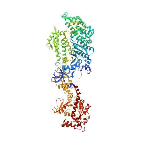
Crystal structure of a dynamin GTPase domain in both nucleotide-free and GDP-bound forms.

Niemann, H.H.Knetsch, M.L.Scherer, A.Manstein, D.J.Kull, F.J.

(2001) EMBO J 20: 5813-5821

  • DOI: https://doi.org/10.1093/emboj/20.21.5813
  • Primary Citation Related Structures: 
    1JWY, 1JX2

  • PubMed Abstract: 

    Dynamins form a family of multidomain GTPases involved in endocytosis, vesicle trafficking and maintenance of mitochondrial morphology. In contrast to the classical switch GTPases, a force-generating function has been suggested for dynamins. Here we report the 2.3 A crystal structure of the nucleotide-free and GDP-bound GTPase domain of Dictyostelium discoideum dynamin A. The GTPase domain is the most highly conserved region among dynamins. The globular structure contains the G-protein core fold, which is extended from a six-stranded beta-sheet to an eight-stranded one by a 55 amino acid insertion. This topologically unique insertion distinguishes dynamins from other subfamilies of GTP-binding proteins. An additional N-terminal helix interacts with the C-terminal helix of the GTPase domain, forming a hydrophobic groove, which could be occupied by C-terminal parts of dynamin not present in our construct. The lack of major conformational changes between the nucleotide-free and the GDP-bound state suggests that mechanochemical rearrangements in dynamin occur during GTP binding, GTP hydrolysis or phosphate release and are not linked to loss of GDP.


  • Organizational Affiliation
    • Department of Biophysics, Max-Planck-Institute for Medical Research, Jahnstrasse 29, D-69120 Heidelberg, Germany.

Macromolecule Content 

  • Total Structure Weight: 125.21 kDa 
  • Atom Count: 8,713 
  • Modeled Residue Count: 1,039 
  • Deposited Residue Count: 1,100 
  • Unique protein chains: 1

Macromolecules

Find similar proteins by:|  3D Structure
Entity ID: 1
MoleculeChains  Sequence LengthOrganismDetailsImage
Myosin-2 heavy chain,Dynamin-A1,100Dictyostelium discoideumMutation(s): 0 
Gene Names: mhcADDB_G0286355dymADDB_G0277849
UniProt
Find proteins for P08799 (Dictyostelium discoideum)
Explore P08799 
Go to UniProtKB:  P08799
Find proteins for Q94464 (Dictyostelium discoideum)
Explore Q94464 
Go to UniProtKB:  Q94464
Entity Groups
Sequence Clusters30% Identity50% Identity70% Identity90% Identity95% Identity100% Identity
UniProt GroupsP08799Q94464
Sequence Annotations
Expand
Reference Sequence

Experimental Data & Validation

Experimental Data

  • Method: X-RAY DIFFRACTION
  • Resolution: 2.30 Å
  • R-Value Free:  0.255 (Depositor), 0.236 (DCC) 
  • R-Value Work:  0.197 (Depositor) 
  • R-Value Observed: 0.197 (Depositor) 
Space Group: P 1 21 1
Unit Cell:
Length ( Å )Angle ( ˚ )
a = 54.45α = 90
b = 62.04β = 94.79
c = 181.2γ = 90
Software Package:
Software NamePurpose
CNSrefinement
MAR345data collection
XDSdata scaling
CNSphasing

Structure Validation

View Full Validation Report


Ligand Structure Quality Assessment 


Entry History 

Deposition Data

Revision History  (Full details and data files)

  • Version 1.0: 2001-11-07
    Type: Initial release
  • Version 1.1: 2008-04-27
    Changes: Version format compliance
  • Version 1.2: 2011-07-13
    Changes: Version format compliance
  • Version 1.3: 2017-05-31
    Changes: Structure summary
  • Version 1.4: 2017-10-04
    Changes: Refinement description
  • Version 1.5: 2020-07-29
    Type: Remediation
    Reason: Carbohydrate remediation
    Changes: Data collection, Derived calculations, Structure summary
  • Version 1.6: 2023-08-16
    Changes: Data collection, Database references, Refinement description, Structure summary
  • Version 1.7: 2024-10-30
    Changes: Structure summary