1JWI

Crystal Structure of Bitiscetin, a von Willeband Factor-dependent Platelet Aggregation Inducer.


Experimental Data Snapshot

  • Method: X-RAY DIFFRACTION
  • Resolution: 2.00 Å
  • R-Value Free: 0.252 
  • R-Value Work: 0.207 

wwPDB Validation   3D Report Full Report


This is version 1.3 of the entry. See complete history


Literature

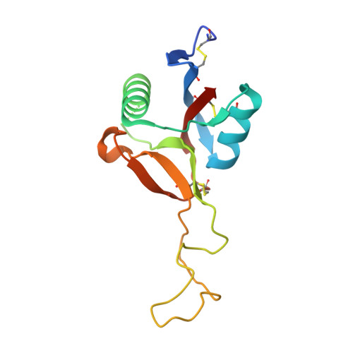
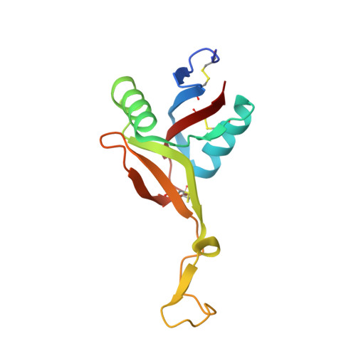
Crystal structure of bitiscetin, a von Willebrand factor-dependent platelet aggregation inducer.

Hirotsu, S.Mizuno, H.Fukuda, K.Qi, M.C.Matsui, T.Hamako, J.Morita, T.Titani, K.

(2001) Biochemistry 40: 13592-13597

  • DOI: https://doi.org/10.1021/bi0114933
  • Primary Citation of Related Structures:  
    1JWI

  • PubMed Abstract: 

    Bitiscetin, a C-type lectin-like protein isolated from the venom of the snake Bitis arientans, promotes the interactions between plasma von Willebrand factor (VWF) and platelet membrane glycoprotein Ib (GPIb) to induce platelet aggregation. We report here the crystal structure of bitiscetin at 2.0 A resolution. The overall fold is similar to those of coagulation factor IX/X-binding protein (IX/X-bp) and flavocetin-A (a GPIb-binding protein), although these three proteins are functionally distinct from one another. The characteristic property determining target recognition is explained mainly by the differences in the surface potential on the central concave surface. A negatively charged patch on the surface of bitiscetin is a candidate for the site of binding to the positively charged surface of the VWF A1 domain, as shown in the case of another platelet aggregation inducer, botrocetin. However, a positively charged patch near the central concave surface is unique for bitiscetin and suggests that it is the binding site for the negatively charged surface of the VWF A3 domain. Thus, the interactions accounting for VWF activation by bitiscetin possibly involve both the A1 and A3 domains of VWF, indicating a specific mechanism of VWF activation by bitiscetin.


  • Organizational Affiliation

    Department of Biochemistry, National Institute of Agrobiological Sciences, Tsukuba, Ibaraki 305-8602, Japan.


Macromolecules
Find similar proteins by:  (by identity cutoff)  |  3D Structure
Entity ID: 1
MoleculeChains Sequence LengthOrganismDetailsImage
bitiscetin131Bitis arietansMutation(s): 0 
UniProt
Find proteins for Q7LZK5 (Bitis arietans)
Explore Q7LZK5 
Go to UniProtKB:  Q7LZK5
Entity Groups  
Sequence Clusters30% Identity50% Identity70% Identity90% Identity95% Identity100% Identity
UniProt GroupQ7LZK5
Sequence Annotations
Expand
  • Reference Sequence
Find similar proteins by:  (by identity cutoff)  |  3D Structure
Entity ID: 2
MoleculeChains Sequence LengthOrganismDetailsImage
platelet aggregation inducer125Bitis arietansMutation(s): 0 
UniProt
Find proteins for Q7LZK8 (Bitis arietans)
Explore Q7LZK8 
Go to UniProtKB:  Q7LZK8
Entity Groups  
Sequence Clusters30% Identity50% Identity70% Identity90% Identity95% Identity100% Identity
UniProt GroupQ7LZK8
Sequence Annotations
Expand
  • Reference Sequence
Experimental Data & Validation

Experimental Data

  • Method: X-RAY DIFFRACTION
  • Resolution: 2.00 Å
  • R-Value Free: 0.252 
  • R-Value Work: 0.207 
  • Space Group: P 21 21 21
Unit Cell:
Length ( Å )Angle ( ˚ )
a = 35.35α = 90
b = 61.74β = 90
c = 120.93γ = 90
Software Package:
Software NamePurpose
DENZOdata reduction
SCALEPACKdata scaling
AMoREphasing
CNSrefinement

Structure Validation

View Full Validation Report


Entry History 

Deposition Data

Revision History  (Full details and data files)

  • Version 1.0: 2001-11-28
    Type: Initial release
  • Version 1.1: 2008-04-27
    Changes: Version format compliance
  • Version 1.2: 2011-07-13
    Changes: Version format compliance
  • Version 1.3: 2023-10-25
    Changes: Data collection, Database references, Refinement description