1JS8

Structure of a Functional Unit from Octopus Hemocyanin


Experimental Data Snapshot

  • Method: X-RAY DIFFRACTION
  • Resolution: 2.30 Å
  • R-Value Free: 0.262 
  • R-Value Work: 0.202 
  • R-Value Observed: 0.201 

wwPDB Validation   3D Report Full Report


Ligand Structure Quality Assessment 


This is version 2.0 of the entry. See complete history


Literature

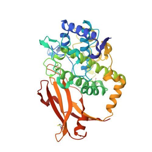
Crystal structure of a functional unit from Octopus hemocyanin.

Cuff, M.E.Miller, K.I.van Holde, K.E.Hendrickson, W.A.

(1998) J Mol Biol 278: 855-870

  • DOI: https://doi.org/10.1006/jmbi.1998.1647
  • Primary Citation of Related Structures:  
    1JS8

  • PubMed Abstract: 

    Hemocyanins are giant oxygen transport proteins found in many arthropods and molluscs. Freely dissolved in the hemolymph, they are multisubunit proteins that contain many copies of the active site, a copper atom pair that reversibly binds oxygen. Octopus hemocyanin is composed of ten subunits, each of which contain seven oxygen-binding "functional units". The carboxyl-terminal 47 kDa functional unit, Odg, is a proteolytic isolate that binds oxygen reversibly while exhibiting slight Bohr and magnesium ion effects. In this work we present the X-ray structure determination and analysis of Odg at 2.3 A resolution. Odg has two structural domains: a largely alpha-helical copper binding domain, and a five-stranded anti-parallel beta-sandwich with the jelly roll topology found in many viruses. Six histidine residues ligate the copper atoms, one of which is involved in a thioether bridge. The results show that the hemocyanin from the mollusc and that from the arthropod have distinct tertiary folds in addition to the long recognized differences in their quaternary structures. Nonetheless, a comparison of Octopus and horseshoe crab hemocyanin reveals a similar active site, in a striking example of perhaps both convergent and divergent evolution.


  • Organizational Affiliation

    Department of Biochemistry and Molecular Biophysics, Columbia University, New York, NY 10032, USA.


Macromolecules
Find similar proteins by:  (by identity cutoff)  |  3D Structure
Entity ID: 1
MoleculeChains Sequence LengthOrganismDetailsImage
Hemocyanin
A, B
394Enteroctopus dofleiniMutation(s): 0 
UniProt
Find proteins for O61363 (Enteroctopus dofleini)
Explore O61363 
Go to UniProtKB:  O61363
Entity Groups  
Sequence Clusters30% Identity50% Identity70% Identity90% Identity95% Identity100% Identity
UniProt GroupO61363
Sequence Annotations
Expand
  • Reference Sequence
Oligosaccharides

Help

Entity ID: 2
MoleculeChains Length2D Diagram Glycosylation3D Interactions
alpha-D-mannopyranose-(1-3)-[alpha-D-mannopyranose-(1-6)]alpha-D-mannopyranose-(1-6)-[beta-D-mannopyranose-(1-3)]beta-D-mannopyranose-(1-4)-2-acetamido-2-deoxy-beta-D-glucopyranose-(1-4)-2-acetamido-2-deoxy-alpha-D-glucopyranose
C
7N/A
Glycosylation Resources
GlyTouCan:  G47457DK
GlyCosmos:  G47457DK
GlyGen:  G47457DK
Entity ID: 3
MoleculeChains Length2D Diagram Glycosylation3D Interactions
2-acetamido-2-deoxy-beta-D-glucopyranose-(1-4)-2-acetamido-2-deoxy-beta-D-glucopyranose
D
2N-Glycosylation
Glycosylation Resources
GlyTouCan:  G42666HT
GlyCosmos:  G42666HT
GlyGen:  G42666HT
Entity ID: 4
MoleculeChains Length2D Diagram Glycosylation3D Interactions
alpha-D-mannopyranose-(1-3)-[beta-D-mannopyranose-(1-6)]alpha-D-mannopyranose
E
3N/A
Glycosylation Resources
GlyTouCan:  G08739AJ
GlyCosmos:  G08739AJ
GlyGen:  G08739AJ
Experimental Data & Validation

Experimental Data

  • Method: X-RAY DIFFRACTION
  • Resolution: 2.30 Å
  • R-Value Free: 0.262 
  • R-Value Work: 0.202 
  • R-Value Observed: 0.201 
  • Space Group: P 21 21 21
Unit Cell:
Length ( Å )Angle ( ˚ )
a = 92.86α = 90
b = 168.39β = 90
c = 58.29γ = 90
Software Package:
Software NamePurpose
DENZOdata reduction
ROTAVATAdata reduction
MADSYSphasing
MLPHAREphasing
X-PLORrefinement
CCP4data scaling
ROTAVATAdata scaling

Structure Validation

View Full Validation Report


Ligand Structure Quality Assessment 


Entry History 

Deposition Data

Revision History  (Full details and data files)

  • Version 1.0: 2001-10-03
    Type: Initial release
  • Version 1.1: 2007-10-16
    Changes: Version format compliance
  • Version 1.2: 2011-07-13
    Changes: Non-polymer description, Version format compliance
  • Version 1.3: 2017-10-04
    Changes: Advisory, Refinement description
  • Version 2.0: 2020-07-29
    Type: Remediation
    Reason: Carbohydrate remediation
    Changes: Advisory, Atomic model, Data collection, Derived calculations, Structure summary