1JD5

Crystal Structure of DIAP1-BIR2/GRIM


Experimental Data Snapshot

  • Method: X-RAY DIFFRACTION
  • Resolution: 1.90 Å
  • R-Value Free: 0.243 
  • R-Value Work: 0.202 
  • R-Value Observed: 0.203 

wwPDB Validation   3D Report Full Report


This is version 1.4 of the entry. See complete history


Literature

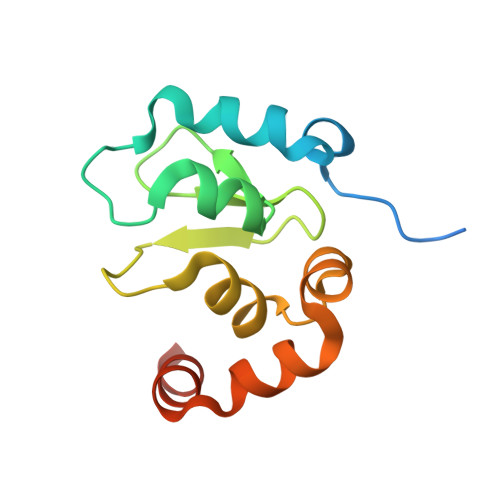
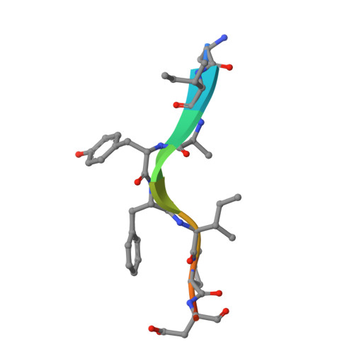
Structural analysis of a functional DIAP1 fragment bound to grim and hid peptides.

Wu, J.W.Cocina, A.E.Chai, J.Hay, B.A.Shi, Y.

(2001) Mol Cell 8: 95-104

  • DOI: https://doi.org/10.1016/s1097-2765(01)00282-9
  • Primary Citation of Related Structures:  
    1JD4, 1JD5, 1JD6

  • PubMed Abstract: 

    The inhibitor of apoptosis protein DIAP1 suppresses apoptosis in Drosophila, with the second BIR domain (BIR2) playing an important role. Three proteins, Hid, Grim, and Reaper, promote apoptosis, in part by binding to DIAP1 through their conserved N-terminal sequences. The crystal structures of DIAP1-BIR2 by itself and in complex with the N-terminal peptides from Hid and Grim reveal that these peptides bind a surface groove on DIAP1, with the first four amino acids mimicking the binding of the Smac tetrapeptide to XIAP. The next 3 residues also contribute to binding through hydrophobic interactions. Interestingly, peptide binding induces the formation of an additional alpha helix in DIAP1. Our study reveals the structural conservation and diversity necessary for the binding of IAPs by the Drosophila Hid/Grim/Reaper and the mammalian Smac proteins.


  • Organizational Affiliation

    Department of Molecular Biology, Lewis Thomas Laboratory, Princeton University, Princeton, NJ 08544, USA.


Macromolecules
Find similar proteins by:  (by identity cutoff)  |  3D Structure
Entity ID: 1
MoleculeChains Sequence LengthOrganismDetailsImage
APOPTOSIS 1 INHIBITOR124Drosophila melanogasterMutation(s): 0 
Gene Names: DIAP1
UniProt
Find proteins for Q24306 (Drosophila melanogaster)
Explore Q24306 
Go to UniProtKB:  Q24306
Entity Groups  
Sequence Clusters30% Identity50% Identity70% Identity90% Identity95% Identity100% Identity
UniProt GroupQ24306
Sequence Annotations
Expand
  • Reference Sequence

Find similar proteins by:  Sequence   |   3D Structure  

Entity ID: 2
MoleculeChains Sequence LengthOrganismDetailsImage
cell death protein GRIM10N/AMutation(s): 0 
UniProt
Find proteins for Q24570 (Drosophila melanogaster)
Explore Q24570 
Go to UniProtKB:  Q24570
Entity Groups  
Sequence Clusters30% Identity50% Identity70% Identity90% Identity95% Identity100% Identity
UniProt GroupQ24570
Sequence Annotations
Expand
  • Reference Sequence
Small Molecules
Ligands 1 Unique
IDChains Name / Formula / InChI Key2D Diagram3D Interactions
ZN
Query on ZN

Download Ideal Coordinates CCD File 
C [auth A]ZINC ION
Zn
PTFCDOFLOPIGGS-UHFFFAOYSA-N
Experimental Data & Validation

Experimental Data

  • Method: X-RAY DIFFRACTION
  • Resolution: 1.90 Å
  • R-Value Free: 0.243 
  • R-Value Work: 0.202 
  • R-Value Observed: 0.203 
  • Space Group: P 65 2 2
Unit Cell:
Length ( Å )Angle ( ˚ )
a = 62.7α = 90
b = 62.7β = 90
c = 130.7γ = 120
Software Package:
Software NamePurpose
AMoREphasing
X-PLORrefinement
DENZOdata reduction
SCALEPACKdata scaling

Structure Validation

View Full Validation Report


Entry History 

Deposition Data

Revision History  (Full details and data files)

  • Version 1.0: 2001-12-05
    Type: Initial release
  • Version 1.1: 2008-04-27
    Changes: Version format compliance
  • Version 1.2: 2011-07-13
    Changes: Version format compliance
  • Version 1.3: 2018-04-04
    Changes: Data collection
  • Version 1.4: 2024-02-07
    Changes: Data collection, Database references, Derived calculations