1J4X

HUMAN VH1-RELATED DUAL-SPECIFICITY PHOSPHATASE C124S MUTANT-PEPTIDE COMPLEX


Experimental Data Snapshot

  • Method: X-RAY DIFFRACTION
  • Resolution: 2.75 Å
  • R-Value Free: 0.260 
  • R-Value Work: 0.188 
  • R-Value Observed: 0.192 

wwPDB Validation   3D Report Full Report


This is version 1.4 of the entry. See complete history


Literature

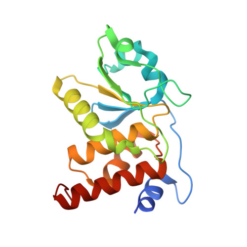
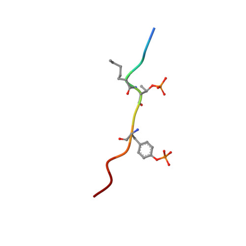
Structural basis for the recognition of a bisphosphorylated MAP kinase peptide by human VHR protein Phosphatase.

Schumacher, M.A.Todd, J.L.Rice, A.E.Tanner, K.G.Denu, J.M.

(2002) Biochemistry 41: 3009-3017

  • DOI: https://doi.org/10.1021/bi015799l
  • Primary Citation of Related Structures:  
    1J4X

  • PubMed Abstract: 

    Human VHR (vaccinia H1 related phosphatase) is a member of the dual-specificity phosphatases (DSPs) that often act on bisphosphorylated protein substrates. Unlike most DSPs, VHR displays a strong preference for dephosphorylating phosphotyrosine residues over phosphothreonine residues. Here we describe the 2.75 A crystal structure of the C124S inactive VHR mutant in complex with a bisphosphorylated peptide corresponding to the MAP kinase activation lip. This structure and subsequent biochemical studies revealed the basis for the strong preference for hydrolyzing phosphotyrosine within bisphosphorylated substrates containing -pTXpY-. In the structure, the two phospho residues are oriented into distinct pockets; the phosphotyrosine is bound in the exposed yet deep active site cleft while the phosphothreonine is loosely tethered into a nearby basic pocket containing Arg(158). As this structure is the first substrate-enzyme complex reported for the DSP family of enzymes, these results provide the first glimpse into how DSPs bind their protein substrates.


  • Organizational Affiliation

    Department of Biochemistry and Molecular Biology, Oregon Health and Science University, Portland, Oregon 97201-3098, USA. schumacn@ohsu.edu


Macromolecules
Find similar proteins by:  (by identity cutoff)  |  3D Structure
Entity ID: 1
MoleculeChains Sequence LengthOrganismDetailsImage
DUAL SPECIFICITY PROTEIN PHOSPHATASE 3184Homo sapiensMutation(s): 1 
EC: 3.1.3.48
UniProt & NIH Common Fund Data Resources
Find proteins for P51452 (Homo sapiens)
Explore P51452 
Go to UniProtKB:  P51452
PHAROS:  P51452
GTEx:  ENSG00000108861 
Entity Groups  
Sequence Clusters30% Identity50% Identity70% Identity90% Identity95% Identity100% Identity
UniProt GroupP51452
Sequence Annotations
Expand
  • Reference Sequence

Find similar proteins by:  Sequence   |   3D Structure  

Entity ID: 2
MoleculeChains Sequence LengthOrganismDetailsImage
DDE(AHP)(TPO)G(PTR)VATRB [auth D]11N/AMutation(s): 0 
Entity Groups  
Sequence Clusters30% Identity50% Identity70% Identity90% Identity95% Identity100% Identity
Sequence Annotations
Expand
  • Reference Sequence
Small Molecules
Modified Residues  3 Unique
IDChains TypeFormula2D DiagramParent
AHP
Query on AHP
B [auth D]L-PEPTIDE LINKINGC7 H15 N O2ALA
PTR
Query on PTR
B [auth D]L-PEPTIDE LINKINGC9 H12 N O6 PTYR
TPO
Query on TPO
B [auth D]L-PEPTIDE LINKINGC4 H10 N O6 PTHR
Experimental Data & Validation

Experimental Data

  • Method: X-RAY DIFFRACTION
  • Resolution: 2.75 Å
  • R-Value Free: 0.260 
  • R-Value Work: 0.188 
  • R-Value Observed: 0.192 
  • Space Group: P 21 21 21
Unit Cell:
Length ( Å )Angle ( ˚ )
a = 34.39α = 90
b = 55.77β = 90
c = 101.13γ = 90
Software Package:
Software NamePurpose
MERLOTphasing
TNTrefinement
MOSFLMdata reduction
CCP4data scaling

Structure Validation

View Full Validation Report


Entry History 

Deposition Data

Revision History  (Full details and data files)

  • Version 1.0: 2001-12-19
    Type: Initial release
  • Version 1.1: 2008-04-26
    Changes: Version format compliance
  • Version 1.2: 2011-07-13
    Changes: Version format compliance
  • Version 1.3: 2021-11-10
    Changes: Database references, Derived calculations
  • Version 1.4: 2023-12-27
    Changes: Data collection