1IRA

COMPLEX OF THE INTERLEUKIN-1 RECEPTOR WITH THE INTERLEUKIN-1 RECEPTOR ANTAGONIST (IL1RA)


Experimental Data Snapshot

  • Method: X-RAY DIFFRACTION
  • Resolution: 2.70 Å
  • R-Value Free: 0.314 
  • R-Value Work: 0.213 
  • R-Value Observed: 0.213 

Starting Models: experimental
View more details

wwPDB Validation   3D Report Full Report


Ligand Structure Quality Assessment 


This is version 1.5 of the entry. See complete history


Literature

A new cytokine-receptor binding mode revealed by the crystal structure of the IL-1 receptor with an antagonist.

Schreuder, H.Tardif, C.Trump-Kallmeyer, S.Soffientini, A.Sarubbi, E.Akeson, A.Bowlin, T.Yanofsky, S.Barrett, R.W.

(1997) Nature 386: 194-200

  • DOI: https://doi.org/10.1038/386194a0
  • Primary Citation of Related Structures:  
    1IRA

  • PubMed Abstract: 

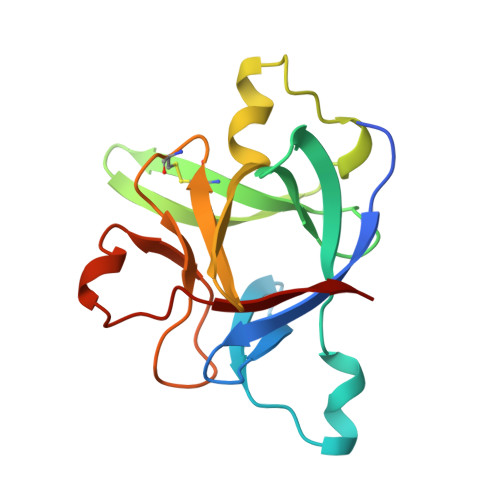
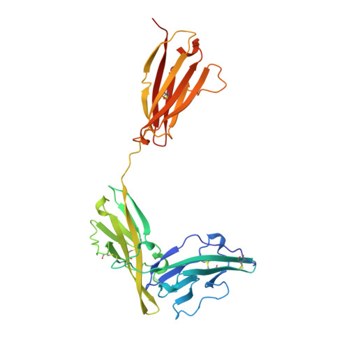
    Inflammation, regardless of whether it is provoked by infection or by tissue damage, starts with the activation of macrophages which initiate a cascade of inflammatory responses by producing the cytokines interleukin-1 (IL-1) and tumour necrosis factor-alpha (ref. 1). Three naturally occurring ligands for the IL-1 receptor (IL1R) exist: the agonists IL-1alpha and IL-1beta and the IL-1-receptor antagonist IL1RA (ref. 2). IL-1 is the only cytokine for which a naturally occurring antagonist is known. Here we describe the crystal structure at 2.7 A resolution of the soluble extracellular part of type-I IL1R complexed with IL1RA. The receptor consists of three immunoglobulin-like domains. Domains 1 and 2 are tightly linked, but domain three is completely separate and connected by a flexible linker. Residues of all three domains contact the antagonist and include the five critical IL1RA residues which were identified by site-directed mutagenesis. A region that is important for biological function in IL-1beta, the 'receptor trigger site' is not in direct contact with the receptor in the IL1RA complex. Modelling studies suggest that this IL-1beta trigger site might induce a movement of domain 3.


  • Organizational Affiliation

    Marion Merrell Dow Research Institute, Strasbourg, France.


Macromolecules
Find similar proteins by:  (by identity cutoff)  |  3D Structure
Entity ID: 1
MoleculeChains Sequence LengthOrganismDetailsImage
INTERLEUKIN-1 RECEPTOR ANTAGONISTA [auth X]152Homo sapiensMutation(s): 0 
UniProt & NIH Common Fund Data Resources
Find proteins for P18510 (Homo sapiens)
Explore P18510 
Go to UniProtKB:  P18510
PHAROS:  P18510
GTEx:  ENSG00000136689 
Entity Groups  
Sequence Clusters30% Identity50% Identity70% Identity90% Identity95% Identity100% Identity
UniProt GroupP18510
Sequence Annotations
Expand
  • Reference Sequence
Find similar proteins by:  (by identity cutoff)  |  3D Structure
Entity ID: 2
MoleculeChains Sequence LengthOrganismDetailsImage
INTERLEUKIN-1 RECEPTORB [auth Y]319Homo sapiensMutation(s): 0 
UniProt & NIH Common Fund Data Resources
Find proteins for P14778 (Homo sapiens)
Explore P14778 
Go to UniProtKB:  P14778
PHAROS:  P14778
GTEx:  ENSG00000115594 
Entity Groups  
Sequence Clusters30% Identity50% Identity70% Identity90% Identity95% Identity100% Identity
UniProt GroupP14778
Glycosylation
Glycosylation Sites: 4Go to GlyGen: P14778-1
Sequence Annotations
Expand
  • Reference Sequence
Experimental Data & Validation

Experimental Data

  • Method: X-RAY DIFFRACTION
  • Resolution: 2.70 Å
  • R-Value Free: 0.314 
  • R-Value Work: 0.213 
  • R-Value Observed: 0.213 
  • Space Group: P 21 21 21
Unit Cell:
Length ( Å )Angle ( ˚ )
a = 47.2α = 90
b = 84.6β = 90
c = 140.2γ = 90
Software Package:
Software NamePurpose
XDSdata scaling
XSCALEdata scaling
SQUASHphasing
X-PLORmodel building
X-PLORrefinement
XDSdata reduction
X-PLOR3.1phasing

Structure Validation

View Full Validation Report


Ligand Structure Quality Assessment 


Entry History 

Deposition Data

Revision History  (Full details and data files)

  • Version 1.0: 1998-06-17
    Type: Initial release
  • Version 1.1: 2008-03-24
    Changes: Version format compliance
  • Version 1.2: 2011-07-13
    Changes: Non-polymer description, Version format compliance
  • Version 1.3: 2018-04-04
    Changes: Data collection
  • Version 1.4: 2020-07-29
    Type: Remediation
    Reason: Carbohydrate remediation
    Changes: Data collection, Derived calculations, Structure summary
  • Version 1.5: 2023-08-09
    Changes: Database references, Refinement description, Structure summary