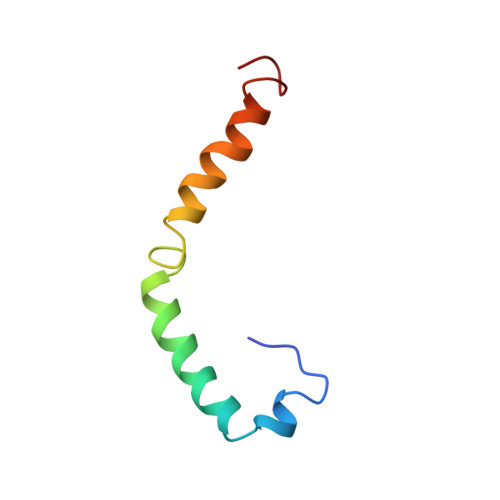
1IOJ

HUMAN APOLIPOPROTEIN C-I, NMR, 18 STRUCTURES


Experimental Data Snapshot

  • Method: SOLUTION NMR
  • Conformers Calculated: 50 
  • Conformers Submitted: 18 
  • Selection Criteria: SELECTED STRUCTURES HAVE NO HELIX-HELIX CONTACTS <5 ANGSTROMS 

wwPDB Validation   3D Report Full Report


This is version 1.3 of the entry. See complete history


Literature

Conformation of human apolipoprotein C-I in a lipid-mimetic environment determined by CD and NMR spectroscopy.

Rozek, A.Sparrow, J.T.Weisgraber, K.H.Cushley, R.J.

(1999) Biochemistry 38: 14475-14484

  • DOI: https://doi.org/10.1021/bi982966h
  • Primary Citation of Related Structures:  
    1IOJ

  • PubMed Abstract: 
  • The high-resolution conformation of human apoC-I in complexes with sodium dodecyl sulfate (SDS) is presented. As estimated from CD data, apoC-I adopts 54% helical secondary structure when bound to SDS, which is similar to the helical content previously found with phospholipids ...

    The high-resolution conformation of human apoC-I in complexes with sodium dodecyl sulfate (SDS) is presented. As estimated from CD data, apoC-I adopts 54% helical secondary structure when bound to SDS, which is similar to the helical content previously found with phospholipids. The NMR-derived conformation of apoC-I is composed of two amphipathic helices, residues 7-29 and 38-52, separated by a flexible linker. The N-terminal helix contains a mobile hinge involving residues 12-15. The hydrophobic side chains cluster on the nonpolar face of both helices, thus forming two discrete lipid-binding sites in the N-terminal helix and one in the C-terminal helix. As suggested by amide proton resonance line widths and deuterium exchange rates, the N-terminal helix is more flexible and may bind less tightly to the detergent than the C-terminal helix. The different mobility of both helices appears to be related to side-chain composition, rather than length of the amphipathic helix, and may play a role in the function of apoC-I as an activator of lecithin:cholesterol acyltransferase (LCAT). A model is suggested in which the C-terminal helix serves as a lipid anchor while the N-terminal helix may hinge off the lipid surface to make specific contacts with LCAT.


    Organizational Affiliation

    Department of Chemistry and Institute of Molecular Biology and Biochemistry, Simon Fraser University, Burnaby, British Columbia Canada.


Macromolecules
Find similar proteins by:  (by identity cutoff)  |  3D Structure
Entity ID: 1
MoleculeChainsSequence LengthOrganismDetailsImage
APOC-I57Homo sapiensMutation(s): 0 
Gene Names: APOC1
Membrane Entity: Yes 
UniProt & NIH Common Fund Data Resources
Find proteins for P02654 (Homo sapiens)
Explore P02654 
Go to UniProtKB:  P02654
PHAROS:  P02654
Entity Groups  
Sequence Clusters30% Identity50% Identity70% Identity90% Identity95% Identity100% Identity
UniProt GroupP02654
Protein Feature View
Expand
  • Reference Sequence
Experimental Data & Validation

Experimental Data

  • Method: SOLUTION NMR
  • Conformers Calculated: 50 
  • Conformers Submitted: 18 
  • Selection Criteria: SELECTED STRUCTURES HAVE NO HELIX-HELIX CONTACTS <5 ANGSTROMS 

Structure Validation

View Full Validation Report


Entry History 

Deposition Data

Revision History  (Full details and data files)

  • Version 1.0: 1998-08-12
    Type: Initial release
  • Version 1.1: 2008-03-04
    Changes: Version format compliance
  • Version 1.2: 2011-07-13
    Changes: Version format compliance
  • Version 1.3: 2018-03-14
    Changes: Data collection, Derived calculations, Experimental preparation, Other, Refinement description, Source and taxonomy, Structure summary