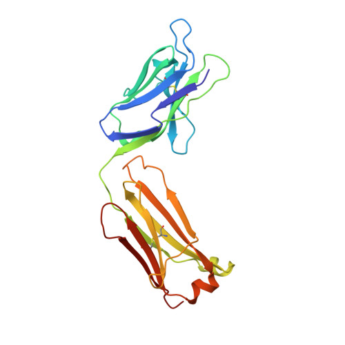
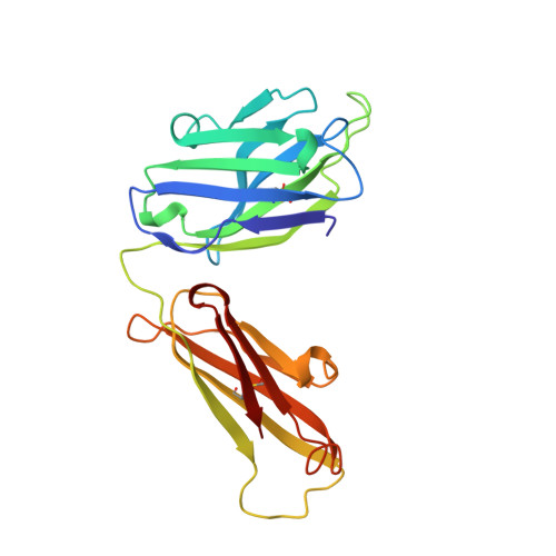
Structure and Specificity of the Anti-Digoxin Antibody 40-50
Jeffrey, P.D., Schildbach, J.F., Chang, C.Y., Kussie, P.H., Margolies, M.N., Sheriff, S.(1995) J Mol Biology 248: 344-360
- PubMed: 7739045 Search on PubMed
- DOI: https://doi.org/10.1016/s0022-2836(95)80055-7
- Primary Citation Related Structures:
1IBG - PubMed Abstract:
We determined the sequence, specificity for structurally related cardenolides, and three-dimensional structure of the anti-digoxin antibody 40-50 Fab in complex with ouabain. The 40-50 antibody does not share close sequence homology with other high-affinity anti-digoxin antibodies. Measurement of the binding constants of structurally distinct digoxin analogs indicated a well-defined specificity pattern also distinct from other anti-digoxin antibodies. The 40-50-ouabain Fab complex crystallizes in space group C2 with cell dimensions of a = 93.7 A, b = 84.8 A, c = 70.1 A, beta = 128.0 degrees. The structure of the complex was determined by X-ray crystallography and refined at a resolution of 2.7 A. The hapten is bound in a pocket extending as a groove from the center of the combining site across the light chain variable domain, with five of the six complementarity-determining regions involved in interactions with the hapten. Approximately three-quarters of the hapten surface area is buried in the complex; two hydrogen bonds are formed between the antibody and hapten. The surface area of the antibody combining site buried by ouabain is contributed equally by the light and heavy chain variable domains. Over half of the surface area buried on the Fab consists of the aromatic side-chains. The surface complementarity between hapten and antibody is sufficient to make the complex specific for only one lactone ring conformation in the hapten. The crystal structure of the 40-50-ouabain complex allows qualitative explanation of the observed fine specificities of 40-50, including that for the binding of haptens substituted at the 16 and 12 positions. Comparison of the crystal structures of 40-50 complexed with ouabain and the previously determined 26-10 anti-digoxin Fab complexed with digoxin, demonstrates that the antibodies bind these structurally related haptens in different orientations, consistent with their different fine specificities. These results demonstrate that the immune system can generate antibodies that provide diverse structural solutions to the binding of even small molecules.
- Department of Macromolecular Crystallography, Bristol-Myers Squibb Pharmaceutical Research Institute, Princeton, NJ 08543-4000, USA.
Organizational Affiliation: