Structural and dynamic perturbations induced by heme binding in cytochrome b5.
Falzone, C.J., Wang, Y., Vu, B.C., Scott, N.L., Bhattacharya, S., Lecomte, J.T.(2001) Biochemistry 40: 4879-4891
- PubMed: 11294656 Search on PubMed
- DOI: https://doi.org/10.1021/bi002681g
- Primary Citation Related Structures:
1I87, 1I8C - PubMed Abstract:
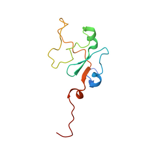
The water-soluble domain of rat hepatic cytochrome b(5) undergoes marked structural changes upon heme removal. The solution structure of apocytochrome b(5) shows that the protein is partially folded in the absence of the heme group, exhibiting a stable module and a disordered heme-binding loop. The quality of the apoprotein structure in solution was improved with the use of heteronuclear NMR data. Backbone amide hydrogen exchange was studied to characterize cooperative units in the protein. It was found that this criterion distinguished the folded module from the heme-binding loop in the apoprotein, in contrast to the holoprotein. The osmolyte trimethylamine N-oxide (TMAO) did not affect the structure of the apoprotein in the disordered region. TMAO imparted a small stabilization consistent with an unfolded state effect correlating with the extent of buried surface area in the folded region of the native apoprotein. The failure of the osmolyte to cause large conformational shifts in the disordered loop supported the view that the specificity of the local sequence for the holoprotein fold was best developed with the stabilization of the native state through heme binding. To dissect the role of the heme prosthetic group in forcing the disordered region into the holoprotein conformation, the axial histidine belonging to the flexible loop (His63) was replaced with an alanine, and the structural properties of the protein with carbon-monoxide-ligated reduced iron were studied. The His63Ala substitution resulted in a protein with lower heme affinity but nevertheless capable of complete refolding. This indicated that the coordination bond was not necessary to establish the structural features of the holoprotein. In addition, the weak binding of the heme in this protein resulted in conformational shifts at a location distant from the binding site. The data suggested an uneven distribution of cooperative elements in the structure of the cytochrome.
- Department of Chemistry, The Pennsylvania State University, University Park, Pennsylvania 16802, USA.
Organizational Affiliation: