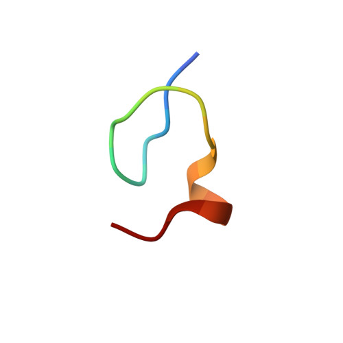
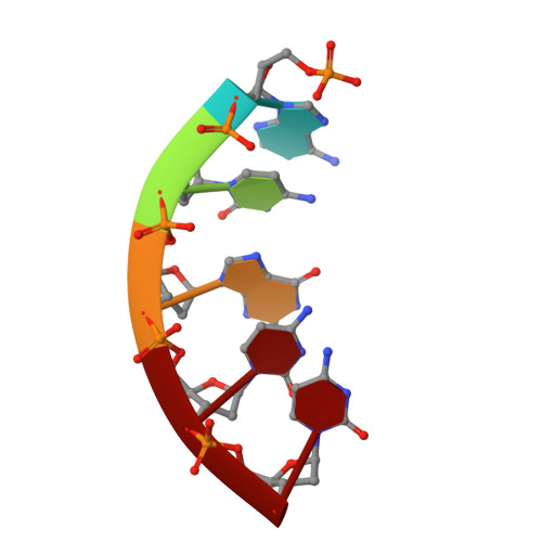
Zinc- and sequence-dependent binding to nucleic acids by the N-terminal zinc finger of the HIV-1 nucleocapsid protein: NMR structure of the complex with the Psi-site analog, dACGCC.
South, T.L., Summers, M.F.(1993) Protein Sci 2: 3
- PubMed: 8443588 Search on PubMedSearch on PubMed Central
- DOI: https://doi.org/10.1002/pro.5560020102
- Primary Citation Related Structures:
1HVN, 1HVO - PubMed Abstract:
The nucleic acid interactive properties of a synthetic peptide with sequence of the N-terminal CCHC zinc finger (CCHC = Cys-X2-Cys-X4-His-X4-Cys; X = variable amino acid) of the human immunodeficiency virus (HIV) nucleocapsid protein, Zn(HIV1-F1), have been studied by 1H NMR spectroscopy. Titration of Zn(HIV1-F1) with oligodeoxyribonucleic acids containing different nucleotide sequences reveals, for the first time, sequence-dependent binding that requires the presence of at least one guanosine residue for tight complex formation. The dynamics of complex formation are sensitive to the nature of the residues adjacent to guanosine, with residues on the 3' side of guanosine having the largest influence. An oligodeoxyribonucleotide with sequence corresponding to a portion of the HIV-1 psi-packaging signal, d(ACGCC), forms a relatively tight complex with Zn(HIV1-F1) (Kd = 5 x 10(-6) M). Two-dimensional nuclear Overhauser effect (NOESY) data indicate that the bound nucleic acid exists predominantly in a single-stranded, A-helical conformation, and the presence of more than a dozen intermolecular NOE cross peaks enabled three-dimensional modeling of the complex. The nucleic acid binds within a hydrophobic cleft on the peptide surface. This hydrophobic cleft is defined by the side chains of residues Val1, Phe4, Ile12, and Ala13. Backbone amide protons of Phe4 and Ala13 and the backbone carbonyl oxygen of Lys2 that lie within this cleft appear to form hydrogen bonds with the guanosine O6 and N1H atoms, respectively. In addition, the positively charged side chain of Arg14 is ideally positioned for electrostatic interactions with the phosphodiester backbone of the nucleic acid. The structural findings provide a rationalization for the general conservation of these hydrophobic and basic residues in CCHC zinc fingers, and are consistent with site-directed mutagenesis results that implicate these residues as direct participants in viral genome recognition.
- Department of Chemistry and Biochemistry, University of Maryland Baltimore County 21228.
Organizational Affiliation: