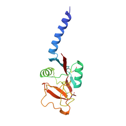
1HUP

HUMAN MANNOSE BINDING PROTEIN CARBOHYDRATE RECOGNITION DOMAIN TRIMERIZES THROUGH A TRIPLE ALPHA-HELICAL COILED-COIL


Experimental Data Snapshot

  • Method: X-RAY DIFFRACTION
  • Resolution: 2.50 Å
  • R-Value Work: 0.199 

wwPDB Validation   3D Report Full Report


This is version 2.0 of the entry. See complete history


Literature

Human mannose-binding protein carbohydrate recognition domain trimerizes through a triple alpha-helical coiled-coil.

Sheriff, S.Chang, C.Y.Ezekowitz, R.A.

(1994) Nat Struct Biol 1: 789-794

  • DOI: https://doi.org/10.1038/nsb1194-789
  • Primary Citation of Related Structures:  
    1HUP

  • PubMed Abstract: 

    Human mannose-binding protein is a hexamer of trimers with each subunit consisting of an amino-terminal region rich in cysteine, 19 collagen repeats, a 'neck', and a carbohydrate recognition domain that requires calcium to bind ligand. A 148-residue peptide, consisting of the 'neck' and carbohydrate recognition domains forms trimers in solution and in crystals. The structure of this trimeric peptide has been determined in two different crystal forms. The 'neck' forms a triple alpha-helical coiled-coil. Each alpha-helix interacts with a neighbouring carbohydrate recognition domain. The spatial arrangement of the carbohydrate recognition domains suggest how MBP trimers form the basic recognition unit for branched oligosaccharides on microorganisms.


  • Organizational Affiliation

    Bristol-Myers Squibb Pharmaceutical Research Institute, Princeton, NJ 08543-4000, USA.


Macromolecules
Find similar proteins by:  (by identity cutoff)  |  3D Structure
Entity ID: 1
MoleculeChains Sequence LengthOrganismDetailsImage
MANNOSE-BINDING PROTEIN141Homo sapiensMutation(s): 0 
UniProt & NIH Common Fund Data Resources
Find proteins for P11226 (Homo sapiens)
Explore P11226 
Go to UniProtKB:  P11226
PHAROS:  P11226
GTEx:  ENSG00000165471 
Entity Groups  
Sequence Clusters30% Identity50% Identity70% Identity90% Identity95% Identity100% Identity
UniProt GroupP11226
Sequence Annotations
Expand
  • Reference Sequence
Experimental Data & Validation

Experimental Data

  • Method: X-RAY DIFFRACTION
  • Resolution: 2.50 Å
  • R-Value Work: 0.199 
  • Space Group: P 3 2 1
Unit Cell:
Length ( Å )Angle ( ˚ )
a = 76.68α = 90
b = 76.68β = 90
c = 58.58γ = 120
Software Package:
Software NamePurpose
X-PLORmodel building
X-PLORrefinement
X-PLORphasing

Structure Validation

View Full Validation Report


Entry History 

Deposition Data

  • Released Date: 1995-10-15 
  • Deposition Author(s): Sheriff, S.

Revision History  (Full details and data files)

  • Version 1.0: 1995-10-15
    Type: Initial release
  • Version 1.1: 2008-03-24
    Changes: Version format compliance
  • Version 1.2: 2011-07-13
    Changes: Derived calculations, Version format compliance
  • Version 1.3: 2015-10-14
    Changes: Refinement description
  • Version 2.0: 2024-06-05
    Changes: Atomic model, Data collection, Database references, Derived calculations, Other