1HJ3 | pdb_00001hj3

Cytochrome cd1 Nitrite Reductase, dioxygen complex


Experimental Data Snapshot

  • Method: X-RAY DIFFRACTION
  • Resolution: 1.60 Å
  • R-Value Free: 
    0.219 (Depositor) 
  • R-Value Work: 
    0.196 (Depositor) 

Starting Model: experimental
View more details

wwPDB Validation 3D Report Full Report

Validation slider image for 1HJ3

This is version 1.6 of the entry. See complete history

Literature

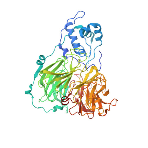
Structure of the bound dioxygen species in the cytochrome oxidase reaction of cytochrome cd1 nitrite reductase.

Sjogren, T.Hajdu, J.

(2001) J Biological Chem 276: 13072-13076

  • DOI: https://doi.org/10.1074/jbc.M011312200
  • Primary Citation Related Structures: 
    1HJ3, 1HJ4, 1HJ5

  • PubMed Abstract: 

    Reduction of dioxygen to water is a key process in aerobic life, but atomic details of this reaction have been elusive because of difficulties in observing active oxygen intermediates by crystallography. Cytochrome cd(1) is a bifunctional enzyme, capable of catalyzing the one-electron reduction of nitrite to nitric oxide, and the four-electron reduction of dioxygen to water. The latter is a cytochrome oxidase reaction. Here we describe the structure of an active dioxygen species in the enzyme captured by cryo-trapping. The productive binding mode of dioxygen in the active site is very similar to that of nitrite and suggests that the catalytic mechanisms of oxygen reduction and nitrite reduction are closely related. This finding has implications to the understanding of the evolution of oxygen-reducing enzymes. Comparison of the dioxygen complex to complexes of cytochrome cd(1) with stable diatomic ligands shows that nitric oxide and cyanide bind in a similar bent conformation to the iron as dioxygen whereas carbon monoxide forms a linear complex. The significance of these differences is discussed.


  • Organizational Affiliation
    • Department of Biochemistry, Uppsala University, Biomedical Center, Box 576, S-751 23 Uppsala, Sweden.

Macromolecule Content 

  • Total Structure Weight: 128.26 kDa 
  • Atom Count: 9,453 
  • Modeled Residue Count: 1,086 
  • Deposited Residue Count: 1,134 
  • Unique protein chains: 1

Macromolecules

Find similar proteins by:|  3D Structure
Entity ID: 1
MoleculeChains  Sequence LengthOrganismDetailsImage
Nitrite reductase
A, B
567Paracoccus pantotrophusMutation(s): 0 
EC: 1.7.2.1 (PDB Primary Data), 1.7.99.1 (PDB Primary Data)
UniProt
Find proteins for P72181 (Paracoccus pantotrophus)
Explore P72181 
Go to UniProtKB:  P72181
Entity Groups
Sequence Clusters30% Identity50% Identity70% Identity90% Identity95% Identity100% Identity
UniProt GroupP72181
Sequence Annotations
Expand
Reference Sequence

Small Molecules

Ligands 5 Unique
IDChains Name / Formula / InChI Key2D Diagram3D Interactions
DHE

Query on DHE


Download:Ideal Coordinates CCD File
D [auth A],
H [auth B]
HEME D
C34 H32 Fe N4 O10
XLQCGNUTSJTZNF-YDXXJHAFSA-L
HEC

Query on HEC


Download:Ideal Coordinates CCD File
C [auth A],
G [auth B]
HEME C
C34 H34 Fe N4 O4
HXQIYSLZKNYNMH-LJNAALQVSA-N
SO4

Query on SO4


Download:Ideal Coordinates CCD File
F [auth A],
K [auth B],
L [auth B]
SULFATE ION
O4 S
QAOWNCQODCNURD-UHFFFAOYSA-L
GOL

Query on GOL


Download:Ideal Coordinates CCD File
E [auth A],
J [auth B]
GLYCEROL
C3 H8 O3
PEDCQBHIVMGVHV-UHFFFAOYSA-N
OXY

Query on OXY


Download:Ideal Coordinates CCD File
I [auth B]OXYGEN MOLECULE
O2
MYMOFIZGZYHOMD-UHFFFAOYSA-N

Experimental Data & Validation

Experimental Data

  • Method: X-RAY DIFFRACTION
  • Resolution: 1.60 Å
  • R-Value Free:  0.219 (Depositor) 
  • R-Value Work:  0.196 (Depositor) 
Space Group: P 1 21 1
Unit Cell:
Length ( Å )Angle ( ˚ )
a = 107.19α = 90
b = 61.17β = 112.19
c = 100.52γ = 90
Software Package:
Software NamePurpose
REFMACrefinement
DENZOdata reduction
SCALEPACKdata scaling

Structure Validation

View Full Validation Report


Entry History 

Deposition Data

Revision History  (Full details and data files)

  • Version 1.0: 2001-01-16
    Type: Initial release
  • Version 1.1: 2011-05-08
    Changes: Version format compliance
  • Version 1.2: 2011-07-13
    Changes: Version format compliance
  • Version 1.3: 2018-06-20
    Changes: Data collection, Database references, Source and taxonomy, Structure summary
  • Version 1.4: 2019-05-08
    Changes: Data collection, Derived calculations, Experimental preparation
  • Version 1.5: 2023-12-13
    Changes: Data collection, Database references, Derived calculations, Other, Refinement description
  • Version 1.6: 2024-11-06
    Changes: Structure summary