1HHP | pdb_00001hhp

THE THREE-DIMENSIONAL STRUCTURE OF THE ASPARTYL PROTEASE FROM THE HIV-1 ISOLATE BRU


Experimental Data Snapshot

  • Method: X-RAY DIFFRACTION
  • Resolution: 2.70 Å
  • R-Value Work: 
    0.190 (Depositor) 

wwPDB Validation 3D Report Full Report

Validation slider image for 1HHP

This is version 1.3 of the entry. See complete history

Literature

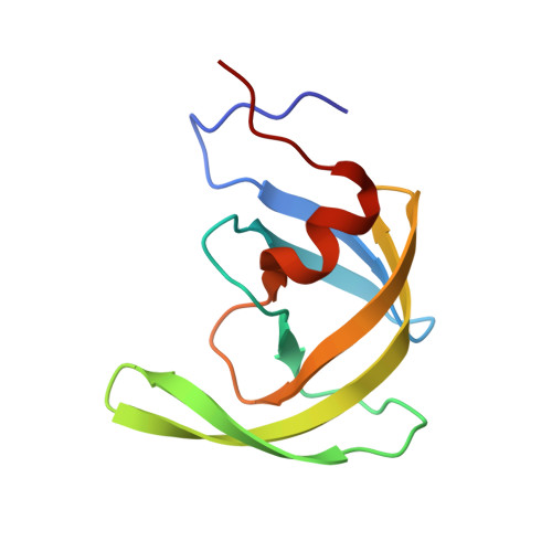
The three-dimensional structure of the aspartyl protease from the HIV-1 isolate BRU.

Spinelli, S.Liu, Q.Z.Alzari, P.M.Hirel, P.H.Poljak, R.J.

(1991) Biochimie 73: 1391-1396

  • DOI: https://doi.org/10.1016/0300-9084(91)90169-2
  • Primary Citation Related Structures: 
    1HHP

  • PubMed Abstract: 

    The crystal structure of the aspartyl protease encoded by the gene pol of the human immunodeficiency virus (HIV-1, isolate BRU) has been determined to 2.7 A resolution. The enzyme, expressed as an insoluble denatured polypeptide in inclusion bodies of Escherichia coli has been renatured and crystallized. It differs by several amino acid replacements from the homologous enzymes of other HIV-1 isolates. A superposition of the C alpha-backbone of the BRU protease with that of the SF2 protease gives a roots mean square positional difference of 0.45 A. Thus, neither the denaturation/renaturation process nor the amino acid replacements have a noticeable effect on the three-dimensional structure of the BRU protease or on the detailed conformation of the catalytic site, which is very similar to that of other aspartyl proteases.


  • Organizational Affiliation
    • URA 359 CNRS, Département d'Immunologie, Institut Pasteur, Paris, France.

Macromolecule Content 

  • Total Structure Weight: 10.8 kDa 
  • Atom Count: 758 
  • Modeled Residue Count: 99 
  • Deposited Residue Count: 99 
  • Unique protein chains: 1

Macromolecules

Find similar proteins by:|  3D Structure
Entity ID: 1
MoleculeChains  Sequence LengthOrganismDetailsImage
UNLIGANDED HIV-1 PROTEASE99Human immunodeficiency virus type 1 (BRU ISOLATE)Mutation(s): 0 
UniProt
Find proteins for P03367 (Human immunodeficiency virus type 1 group M subtype B (isolate BRU/LAI))
Explore P03367 
Go to UniProtKB:  P03367
Entity Groups
Sequence Clusters30% Identity50% Identity70% Identity90% Identity95% Identity100% Identity
UniProt GroupP03367
Sequence Annotations
Expand
Reference Sequence

Experimental Data & Validation

Experimental Data

  • Method: X-RAY DIFFRACTION
  • Resolution: 2.70 Å
  • R-Value Work:  0.190 (Depositor) 
Space Group: P 41 21 2
Unit Cell:
Length ( Å )Angle ( ˚ )
a = 50.2α = 90
b = 50.2β = 90
c = 106.8γ = 90
Software Package:
Software NamePurpose
X-PLORmodel building
X-PLORrefinement
X-PLORphasing

Structure Validation

View Full Validation Report


Entry History 

Deposition Data

Revision History  (Full details and data files)

  • Version 1.0: 1992-10-15
    Type: Initial release
  • Version 1.1: 2008-03-24
    Changes: Version format compliance
  • Version 1.2: 2011-07-13
    Changes: Derived calculations, Version format compliance
  • Version 1.3: 2024-02-07
    Changes: Data collection, Database references, Other