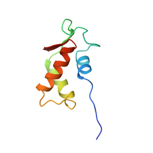
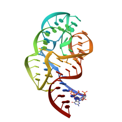
1HC8

CRYSTAL STRUCTURE OF A CONSERVED RIBOSOMAL PROTEIN-RNA COMPLEX


Experimental Data Snapshot

  • Method: X-RAY DIFFRACTION
  • Resolution: 2.80 Å
  • R-Value Free: 0.253 
  • R-Value Work: 0.217 
  • R-Value Observed: 0.217 

wwPDB Validation   3D Report Full Report


This is version 1.2 of the entry. See complete history


Literature

A Compact RNA Tertiary Structure Contains a Buried Backbone-K+ Complex

Conn, G.L.Gittis, A.G.Lattman, E.E.Misra, V.K.Draper, D.E.

(2002) J Mol Biol 318: 963

  • DOI: 10.1016/S0022-2836(02)00147-X
  • Primary Citation of Related Structures:  
    1HC8

  • PubMed Abstract: 
  • The structure of a 58 nucleotide ribosomal RNA fragment buries several phosphate groups of a hairpin loop within a large tertiary core. During refinement of an X-ray crystal structure containing this RNA, a potassium ion was found to be contacted by six oxygen atoms from the buried phosphate groups; the ion is contained completely within the solvent-accessible surface of the RNA ...

    The structure of a 58 nucleotide ribosomal RNA fragment buries several phosphate groups of a hairpin loop within a large tertiary core. During refinement of an X-ray crystal structure containing this RNA, a potassium ion was found to be contacted by six oxygen atoms from the buried phosphate groups; the ion is contained completely within the solvent-accessible surface of the RNA. The electrostatic potential at the ion chelation site is unusually large, and more than compensates for the substantial energetic penalties associated with partial dehydration of the ion and displacement of delocalized ions. The very large predicted binding free energy, approximately -30 kcal/mol, implies that the site must be occupied for the RNA to fold. These findings agree with previous studies of the ion-dependent folding of tertiary structure in this RNA, which concluded that a monovalent ion was bound in a partially dehydrated environment where Mg2+ could not easily compete for binding. By compensating the unfavorable free energy of buried phosphate groups with a chelated ion, the RNA is able to create a larger and more complex tertiary fold than would be possible otherwise.


    Related Citations: 
    • Crystal Structure of a Conserved Ribosomal Protein-RNA Complex
      Conn, G.L., Draper, D.E., Lattman, E.E., Gittis, A.G.
      (1999) Science 284: 1171

    Organizational Affiliation

    Department of Chemistry, The Johns Hopkins University, 3400 North Charles Street, Baltimore, MD 21218, USA.


Macromolecules

Find similar proteins by:  (by identity cutoff)  |  3D Structure
Entity ID: 1
MoleculeChainsSequence LengthOrganismDetailsImage
RIBOSOMAL PROTEIN L11A, B76Geobacillus stearothermophilusMutation(s): 0 
Gene Names: rplK
UniProt
Find proteins for P56210 (Geobacillus stearothermophilus)
Explore P56210 
Go to UniProtKB:  P56210
Entity Groups  
Sequence Clusters30% Identity50% Identity70% Identity90% Identity95% Identity100% Identity
UniProt GroupP56210
Protein Feature View
Expand
  • Reference Sequence
Find similar nucleic acids by:  (by identity cutoff)  |  3D Structure
Entity ID: 2
MoleculeChainsLengthOrganismImage
58 NUCLEOTIDE RIBOSOMAL 23S RNA DOMAINC, D58Escherichia coli
Protein Feature View
Expand
  • Reference Sequence
Small Molecules
Ligands 3 Unique
IDChainsName / Formula / InChI Key2D Diagram3D Interactions
OS
Query on OS

Download Ideal Coordinates CCD File 
CA [auth D],
DA [auth D],
O [auth C],
P [auth C]
OSMIUM ION
Os
XQBKHDFIPARBOX-UHFFFAOYSA-N
 Ligand Interaction
K
Query on K

Download Ideal Coordinates CCD File 
H [auth C],
U [auth D]
POTASSIUM ION
K
NPYPAHLBTDXSSS-UHFFFAOYSA-N
 Ligand Interaction
MG
Query on MG

Download Ideal Coordinates CCD File 
AA [auth D],
BA [auth D],
E [auth C],
EA [auth D],
F [auth C],
AA [auth D],
BA [auth D],
E [auth C],
EA [auth D],
F [auth C],
G [auth C],
I [auth C],
J [auth C],
K [auth C],
L [auth C],
M [auth C],
N [auth C],
Q [auth C],
R [auth D],
S [auth D],
T [auth D],
V [auth D],
W [auth D],
X [auth D],
Y [auth D],
Z [auth D]
MAGNESIUM ION
Mg
JLVVSXFLKOJNIY-UHFFFAOYSA-N
 Ligand Interaction
Experimental Data & Validation

Experimental Data

  • Method: X-RAY DIFFRACTION
  • Resolution: 2.80 Å
  • R-Value Free: 0.253 
  • R-Value Work: 0.217 
  • R-Value Observed: 0.217 
  • Space Group: P 43 21 2
Unit Cell:
Length ( Å )Angle ( ˚ )
a = 150.675α = 90
b = 150.675β = 90
c = 63.841γ = 90
Software Package:
Software NamePurpose
CNSrefinement
DENZOdata reduction
SCALEPACKdata scaling
MLPHAREphasing

Structure Validation

View Full Validation Report


Entry History 

Deposition Data

Revision History  (Full details and data files)

  • Version 1.0: 2002-05-23
    Type: Initial release
  • Version 1.1: 2011-05-08
    Changes: Version format compliance
  • Version 1.2: 2011-07-13
    Changes: Version format compliance