1GVE | pdb_00001gve

Aflatoxin aldehyde reductase (AKR7A1) from Rat Liver


Experimental Data Snapshot

  • Method: X-RAY DIFFRACTION
  • Resolution: 1.38 Å
  • R-Value Free: 
    0.180 (Depositor) 
  • R-Value Work: 
    0.158 (Depositor) 
  • R-Value Observed: 
    0.159 (Depositor) 

Starting Model: experimental
View more details

wwPDB Validation 3D Report Full Report

Validation slider image for 1GVE

This is version 1.4 of the entry. See complete history

Literature

The Crystal Structure of Rat Liver Akr7A1: A Dimeric Member of the Aldo-Keto Reductase Superfamily

Kozma, E.Brown, E.Ellis, E.M.Lapthorn, A.J.

(2002) J Biological Chem 277: 16285

  • DOI: https://doi.org/10.1074/jbc.M110808200
  • Primary Citation Related Structures: 
    1GVE

  • PubMed Abstract: 

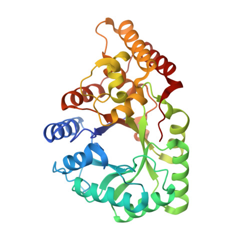
    The structure of the rat liver aflatoxin dialdehyde reductase (AKR7A1) has been solved to 1.38-A resolution. Although it shares a similar alpha/beta-barrel structure with other members of the aldo-keto reductase superfamily, AKR7A1 is the first dimeric member to be crystallized. The crystal structure also reveals details of the ternary complex as one subunit of the dimer contains NADP(+) and the inhibitor citrate. Although the underlying catalytic mechanism appears similar to other aldo-keto reductases, the substrate-binding pocket contains several charged amino acids (Arg-231 and Arg-327) that distinguish it from previously characterized aldo-keto reductases with respect to size and charge. These differences account for the substrate specificity for 4-carbon acid-aldehydes such as succinic semialdehyde and 2-carboxybenzaldehyde as well as for the idiosyncratic substrate aflatoxin B(1) dialdehyde of this subfamily of enzymes. Structural differences between the AKR7A1 ternary complex and apoenzyme reveal a significant hinged movement of the enzyme involving not only the loops of the structure but also parts of the alpha/beta-barrel most intimately involved in cofactor binding.


  • Organizational Affiliation
    • Department of Chemistry, University of Glasgow, Glasgow G12 8QQ, United Kingdom.

Macromolecule Content 

  • Total Structure Weight: 75.62 kDa 
  • Atom Count: 5,567 
  • Modeled Residue Count: 625 
  • Deposited Residue Count: 654 
  • Unique protein chains: 1

Macromolecules

Find similar proteins by:|  3D Structure
Entity ID: 1
MoleculeChains  Sequence LengthOrganismDetailsImage
AFLATOXIN B1 ALDEHYDE REDUCTASE MEMBER 3
A, B
327Rattus norvegicusMutation(s): 0 
EC: 1 (PDB Primary Data), 1.1.1.2 (UniProt)
UniProt
Find proteins for P38918 (Rattus norvegicus)
Explore P38918 
Go to UniProtKB:  P38918
Entity Groups
Sequence Clusters30% Identity50% Identity70% Identity90% Identity95% Identity100% Identity
UniProt GroupP38918
Sequence Annotations
Expand
Reference Sequence

Small Molecules

Ligands 3 Unique
IDChains Name / Formula / InChI Key2D Diagram3D Interactions
NAP

Query on NAP


Download:Ideal Coordinates CCD File
C [auth A]NADP NICOTINAMIDE-ADENINE-DINUCLEOTIDE PHOSPHATE
C21 H28 N7 O17 P3
XJLXINKUBYWONI-NNYOXOHSSA-N
CIT

Query on CIT


Download:Ideal Coordinates CCD File
F [auth A]CITRIC ACID
C6 H8 O7
KRKNYBCHXYNGOX-UHFFFAOYSA-N
GOL

Query on GOL


Download:Ideal Coordinates CCD File
D [auth A]
E [auth A]
G [auth B]
H [auth B]
I [auth B]
D [auth A],
E [auth A],
G [auth B],
H [auth B],
I [auth B],
J [auth B],
K [auth B],
L [auth B],
M [auth B],
N [auth B],
O [auth B],
P [auth B]
GLYCEROL
C3 H8 O3
PEDCQBHIVMGVHV-UHFFFAOYSA-N

Experimental Data & Validation

Experimental Data

  • Method: X-RAY DIFFRACTION
  • Resolution: 1.38 Å
  • R-Value Free:  0.180 (Depositor) 
  • R-Value Work:  0.158 (Depositor) 
  • R-Value Observed: 0.159 (Depositor) 
Space Group: C 1 2 1
Unit Cell:
Length ( Å )Angle ( ˚ )
a = 125.54α = 90
b = 64.68β = 91.03
c = 112.84γ = 90
Software Package:
Software NamePurpose
REFMACrefinement
DENZOdata reduction
SCALEPACKdata scaling
AMoREphasing

Structure Validation

View Full Validation Report


Entry History 

Deposition Data

Revision History  (Full details and data files)

  • Version 1.0: 2002-06-27
    Type: Initial release
  • Version 1.1: 2011-09-21
    Changes: Atomic model, Data collection, Database references, Derived calculations, Non-polymer description, Other, Refinement description, Source and taxonomy, Structure summary, Version format compliance
  • Version 1.2: 2019-05-08
    Changes: Data collection, Experimental preparation, Other
  • Version 1.3: 2019-07-24
    Changes: Data collection
  • Version 1.4: 2023-12-13
    Changes: Data collection, Database references, Derived calculations, Refinement description