1G5N

ANNEXIN V COMPLEX WITH HEPARIN OLIGOSACCHARIDES


Experimental Data Snapshot

  • Method: X-RAY DIFFRACTION
  • Resolution: 1.90 Å
  • R-Value Free: 0.238 
  • R-Value Work: 0.208 
  • R-Value Observed: 0.208 

wwPDB Validation   3D Report Full Report


This is version 2.2 of the entry. See complete history


Literature

Annexin V--heparin oligosaccharide complex suggests heparan sulfate--mediated assembly on cell surfaces.

Capila, I.Hernaiz, M.J.Mo, Y.D.Mealy, T.R.Campos, B.Dedman, J.R.Linhardt, R.J.Seaton, B.A.

(2001) Structure 9: 57-64

  • DOI: https://doi.org/10.1016/s0969-2126(00)00549-9
  • Primary Citation of Related Structures:  
    1G5N

  • PubMed Abstract: 

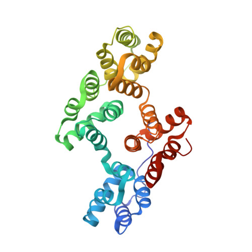
    Annexin V, an abundant anticoagulant protein, has been proposed to exert its effects by self-assembling into highly ordered arrays on phospholipid membranes to form a protective anti-thrombotic shield at the cell surface. The protein exhibits very high-affinity calcium-dependent interactions with acidic phospholipid membranes, as well as specific binding to glycosaminoglycans (GAGs) such as heparin and heparan sulfate, a major component of cell surface proteoglycans. At present, there is no structural information to elucidate this interaction or the role it may play in annexin V function at the cell surface. We report the 1.9 A crystal structure of annexin V in complex with heparin-derived tetrasaccharides. This structure represents the first of a heparin oligosaccharide binding to a protein where calcium ions are essential for the interaction. Two distinct GAG binding sites are situated on opposite protein surfaces. Basic residues at each site were identified from the structure and site-directed mutants were prepared. The heparin binding properties of these mutants were measured by surface plasmon resonance. The results confirm the roles of these mutated residues in heparin binding, and the kinetic and thermodynamic data define the functionally distinct character of each distal binding surface. The annexin V molecule, as it self-assembles into an organized array on the membrane surface, can bind the heparan sulfate components of cell surface proteoglycans. A novel model is presented in which proteoglycan heparan sulfate could assist in the localization of annexin V to the cell surface membrane and/or stabilization of the entire molecular assembly to promote anticoagulation.


  • Organizational Affiliation

    Division of Medicinal and Natural Products Chemistry, Department of Chemistry, University of Iowa, Iowa City, IA 52242, USA.


Macromolecules
Find similar proteins by:  (by identity cutoff)  |  3D Structure
Entity ID: 1
MoleculeChains Sequence LengthOrganismDetailsImage
ANNEXIN V318Rattus norvegicusMutation(s): 0 
Gene Names: LC5
UniProt
Find proteins for P14668 (Rattus norvegicus)
Explore P14668 
Go to UniProtKB:  P14668
Entity Groups  
Sequence Clusters30% Identity50% Identity70% Identity90% Identity95% Identity100% Identity
UniProt GroupP14668
Sequence Annotations
Expand
  • Reference Sequence
Oligosaccharides

Help

Entity ID: 2
MoleculeChains Length2D Diagram Glycosylation3D Interactions
4-deoxy-2-O-sulfo-alpha-L-threo-hex-4-enopyranuronic acid-(1-4)-2-deoxy-6-O-sulfo-2-(sulfoamino)-alpha-D-glucopyranose-(1-4)-2-O-sulfo-alpha-L-idopyranuronic acid-(1-4)-2-deoxy-6-O-sulfo-2-(sulfoamino)-alpha-D-glucopyranose
B, C
4N/A
Glycosylation Resources
GlyTouCan:  G88016IC
GlyCosmos:  G88016IC
Experimental Data & Validation

Experimental Data

  • Method: X-RAY DIFFRACTION
  • Resolution: 1.90 Å
  • R-Value Free: 0.238 
  • R-Value Work: 0.208 
  • R-Value Observed: 0.208 
  • Space Group: H 3
Unit Cell:
Length ( Å )Angle ( ˚ )
a = 155.874α = 90
b = 155.874β = 90
c = 37.344γ = 120
Software Package:
Software NamePurpose
CNSrefinement
DENZOdata reduction
SCALEPACKdata scaling
CNSphasing

Structure Validation

View Full Validation Report


Entry History 

Deposition Data

Revision History  (Full details and data files)

  • Version 1.0: 2001-06-06
    Type: Initial release
  • Version 1.1: 2008-04-27
    Changes: Version format compliance
  • Version 1.2: 2011-07-13
    Changes: Non-polymer description, Version format compliance
  • Version 2.0: 2020-07-29
    Type: Remediation
    Reason: Carbohydrate remediation
    Changes: Atomic model, Data collection, Derived calculations, Structure summary
  • Version 2.1: 2024-02-07
    Changes: Data collection, Database references, Structure summary
  • Version 2.2: 2024-04-03
    Changes: Refinement description