Structure of rat parvalbumin with deleted AB domain: implications for the evolution of EF hand calcium-binding proteins and possible physiological relevance.
Thepaut, M., Strub, M.P., Cave, A., Baneres, J.L., Berchtold, M.W., Dumas, C., Padilla, A.(2001) Proteins 45: 117-128
- PubMed: 11562941 Search on PubMed
- DOI: https://doi.org/10.1002/prot.1131
- Primary Citation Related Structures:
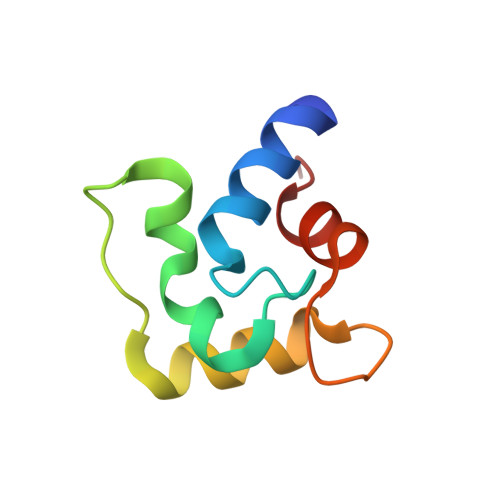
1G33 - PubMed Abstract:
Among the EF-hand Ca(2+)-binding proteins, parvalbumin (PV) and calbindin D9k (CaB) have the function of Ca(2+) buffers. They evolved from an ancestor protein through two phylogenetic pathways, keeping one pair of EF-hands. They differ by the extra helix-loop-helix (AB domain) found in PV and by the linker between the binding sites. To investigate whether the deletion of AB in PV restores a CaB-like structure, we prepared and solved the structure of the truncated rat PV (PVratDelta37) by X-ray and NMR. PVratDelta37 keeps the PV fold, but is more compact, having a well-structured linker, which differs remarkably from CaB. PvratDelta37 has no stable apo-form, has lower affinity for Ca(2+) than full-length PV, and does not bind Mg(2+), in contrast to CaB. Structural differences of the hydrophobic core are partially responsible for lowering the calcium-binding affinity of the truncated protein. It can be concluded that the AB domain, like the linker of CaB, plays a role in structural stabilization. The AB domain of PV protects the hydrophobic core, and is required to maintain high affinity for divalent cation binding. Therefore, the AB domain possibly modulates PV buffer function.
- Centre de Biochimie Structurale, UMR 5048 CNRS, U554 INSERM, Université Montpellier I, Montpellier, France.
Organizational Affiliation: