Structure of ferric soybean leghemoglobin a nicotinate at 2.3 A resolution.
Ellis, P.J., Appleby, C.A., Guss, J.M., Hunter, W.N., Ollis, D.L., Freeman, H.C.(1997) Acta Crystallogr D Biol Crystallogr 53: 302-310
- PubMed: 15299933 Search on PubMed
- DOI: https://doi.org/10.1107/S0907444997000292
- Primary Citation Related Structures:
1FSL - PubMed Abstract:
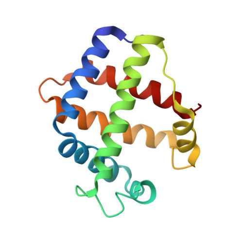
Soybean leghemoglobin a is a small (16 kDa) protein facilitating the transport of O(2) to respiring N(2)-fixing bacteria at low free-O(2) tension. The crystal structure of soybean ferric leghemoglobin a nicotinate has been refined at 2.3 A resolution. The final R factor is 15.8% for 6877 reflections between 6.0 and 2.3 A. The structure of soybean leghemoglobin a (143 residues) is closely similar to that of lupin leghemoglobin II (153 residues), the proteins having 82 identical residues when the sequences are aligned. The new structure provides support for the conclusion that the unique properties of leghemoglobin arise principally from a heme pocket considerably larger and more flexible than that of myoglobin, a strongly ruffled heme group, and a proximal histidine orientation more favourable to ligand binding.
- School of Chemistry, University of Sydney, NSW, Australia.
Organizational Affiliation: