A cluster exposed: structure of the Rieske ferredoxin from biphenyl dioxygenase and the redox properties of Rieske Fe-S proteins.
Colbert, C.L., Couture, M.M., Eltis, L.D., Bolin, J.T.(2000) Structure 8: 1267-1278
- PubMed: 11188691 Search on PubMed
- DOI: https://doi.org/10.1016/s0969-2126(00)00536-0
- Primary Citation Related Structures:
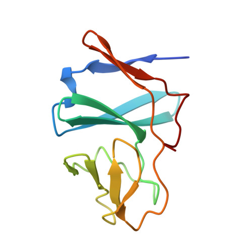
1FQT - PubMed Abstract:
Ring-hydroxylating dioxygenases are multicomponent systems that initiate biodegradation of aromatic compounds. Many dioxygenase systems include Rieske-type ferredoxins with amino acid sequences and redox properties remarkably different from the Rieske proteins of proton-translocating respiratory and photosynthetic complexes. In the latter, the [Fe2S2] clusters lie near the protein surface, operate at potentials above +300 mV at pH 7, and express pH- and ionic strength-dependent redox behavior. The reduction potentials of the dioxygenase ferredoxins are approximately 150 mV and are pH-independent. These distinctions were predicted to arise from differences in the exposure of the cluster and/or interactions of the histidine ligands. The crystal structure of BphF, the Rieske-type ferredoxin associated with biphenyl dioxygenase, was determined by multiwavelength anomalous diffraction and refined at 1.6 A resolution. The structure of BphF was compared with other Rieske proteins at several levels. BphF has the same two-domain fold as other Rieske proteins, but it lacks all insertions that give the others unique structural features. The BphF Fe-S cluster and its histidine ligands are exposed. However, the cluster has a significantly different environment in that five fewer polar groups interact strongly with the cluster sulfide or the cysteinyl ligands. BphF has structural features consistent with a minimal and perhaps archetypical Rieske protein. Variations in redox potentials among Rieske clusters appear to be largely the result of local electrostatic interactions with protein partial charges. Moreover, it appears that the redox-linked ionizations of the Rieske proteins from proton-translocating complexes are also promoted by these electrostatic interactions.
- Department of Biological Sciences, Purdue University, West Lafayette, Indiana 47907, USA.
Organizational Affiliation: