1FLH | pdb_00001flh

CRYSTAL STRUCTURE OF HUMAN UROPEPSIN AT 2.45 A RESOLUTION


Experimental Data Snapshot

  • Method: X-RAY DIFFRACTION
  • Resolution: 2.45 Å
  • R-Value Work: 
    0.161 (Depositor), 0.195 (DCC) 
  • R-Value Observed: 
    0.161 (Depositor) 

Starting Model: experimental
View more details

wwPDB Validation 3D Report Full Report

Validation slider image for 1FLH

This is version 1.4 of the entry. See complete history

Literature

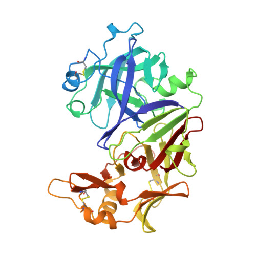
Structure of human uropepsin at 2.45 A resolution.

Canduri, F.Teodoro, L.G.Fadel, V.Lorenzi, C.C.Hial, V.Gomes, R.A.Neto, J.R.de Azevedo, W.F.

(2001) Acta Crystallogr D Biol Crystallogr 57: 1560-1570

  • DOI: https://doi.org/10.1107/s0907444901013865
  • Primary Citation Related Structures: 
    1FLH

  • PubMed Abstract: 

    The molecular structure of human uropepsin, an aspartic proteinase from the urine produced in the form of pepsinogen A in the gastric mucosa, has been determined by molecular replacement using human pepsin as the search model. Crystals belong to space group P2(1)2(1)2(1), with unit-cell parameters a = 50.99, b = 75.56, c = 89.90 A. Crystallographic refinement led to an R factor of 0.161 at 2.45 A resolution. The positions of 2437 non-H protein atoms in 326 residues have been determined and the model contains 143 water molecules. The structure is bilobal, consisting of two predominantly beta-sheet lobes which, as observed in other aspartic proteinases, are related by a pseudo-twofold axis. A model of the uropepsin-pepstatin complex has been constructed based on the high-resolution crystal structure of pepsin complexed with pepstatin.


  • Organizational Affiliation
    • Departamento de Física, IBILCE, UNESP, São José do Rio Preto, SP 15054-000, Brazil.

Macromolecule Content 

  • Total Structure Weight: 34.62 kDa 
  • Atom Count: 2,580 
  • Modeled Residue Count: 326 
  • Deposited Residue Count: 326 
  • Unique protein chains: 1

Macromolecules

Find similar proteins by:|  3D Structure
Entity ID: 1
MoleculeChains  Sequence LengthOrganismDetailsImage
UROPEPSIN326Homo sapiensMutation(s): 0 
EC: 3.4.23.1
UniProt & NIH Common Fund Data Resources
Find proteins for P0DJD7 (Homo sapiens)
Explore P0DJD7 
Go to UniProtKB:  P0DJD7
PHAROS:  P0DJD7
GTEx:  ENSG00000229183 
Entity Groups
Sequence Clusters30% Identity50% Identity70% Identity90% Identity95% Identity100% Identity
UniProt GroupP0DJD7
Sequence Annotations
Expand
Reference Sequence

Experimental Data & Validation

Experimental Data

  • Method: X-RAY DIFFRACTION
  • Resolution: 2.45 Å
  • R-Value Work:  0.161 (Depositor), 0.195 (DCC) 
  • R-Value Observed: 0.161 (Depositor) 
Space Group: P 21 21 21
Unit Cell:
Length ( Å )Angle ( ˚ )
a = 50.989α = 90
b = 75.557β = 90
c = 89.896γ = 90
Software Package:
Software NamePurpose
DENZOdata reduction
SCALEPACKdata scaling
AMoREphasing
X-PLORrefinement

Structure Validation

View Full Validation Report


Entry History 

Deposition Data

Revision History  (Full details and data files)

  • Version 1.0: 2001-10-31
    Type: Initial release
  • Version 1.1: 2008-04-27
    Changes: Version format compliance
  • Version 1.2: 2011-07-13
    Changes: Version format compliance
  • Version 1.3: 2023-08-09
    Changes: Data collection, Database references, Refinement description
  • Version 1.4: 2024-10-09
    Changes: Structure summary