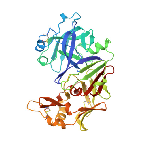
1FLH

CRYSTAL STRUCTURE OF HUMAN UROPEPSIN AT 2.45 A RESOLUTION


Experimental Data Snapshot

  • Method: X-RAY DIFFRACTION
  • Resolution: 2.45 Å
  • R-Value Work: 0.161 
  • R-Value Observed: 0.161 

Starting Model: experimental
View more details

wwPDB Validation   3D Report Full Report


This is version 1.3 of the entry. See complete history


Literature

Structure of human uropepsin at 2.45 A resolution.

Canduri, F.Teodoro, L.G.Fadel, V.Lorenzi, C.C.Hial, V.Gomes, R.A.Neto, J.R.de Azevedo, W.F.

(2001) Acta Crystallogr D Biol Crystallogr 57: 1560-1570

  • DOI: https://doi.org/10.1107/s0907444901013865
  • Primary Citation of Related Structures:  
    1FLH

  • PubMed Abstract: 

    The molecular structure of human uropepsin, an aspartic proteinase from the urine produced in the form of pepsinogen A in the gastric mucosa, has been determined by molecular replacement using human pepsin as the search model. Crystals belong to space group P2(1)2(1)2(1), with unit-cell parameters a = 50.99, b = 75.56, c = 89.90 A. Crystallographic refinement led to an R factor of 0.161 at 2.45 A resolution. The positions of 2437 non-H protein atoms in 326 residues have been determined and the model contains 143 water molecules. The structure is bilobal, consisting of two predominantly beta-sheet lobes which, as observed in other aspartic proteinases, are related by a pseudo-twofold axis. A model of the uropepsin-pepstatin complex has been constructed based on the high-resolution crystal structure of pepsin complexed with pepstatin.


  • Organizational Affiliation

    Departamento de Física, IBILCE, UNESP, São José do Rio Preto, SP 15054-000, Brazil.


Macromolecules
Find similar proteins by:  (by identity cutoff)  |  3D Structure
Entity ID: 1
MoleculeChains Sequence LengthOrganismDetailsImage
UROPEPSIN326Homo sapiensMutation(s): 0 
EC: 3.4.23.1
UniProt & NIH Common Fund Data Resources
Find proteins for P0DJD7 (Homo sapiens)
Explore P0DJD7 
Go to UniProtKB:  P0DJD7
PHAROS:  P0DJD7
GTEx:  ENSG00000229183 
Entity Groups  
Sequence Clusters30% Identity50% Identity70% Identity90% Identity95% Identity100% Identity
UniProt GroupP0DJD7
Sequence Annotations
Expand
  • Reference Sequence
Experimental Data & Validation

Experimental Data

  • Method: X-RAY DIFFRACTION
  • Resolution: 2.45 Å
  • R-Value Work: 0.161 
  • R-Value Observed: 0.161 
  • Space Group: P 21 21 21
Unit Cell:
Length ( Å )Angle ( ˚ )
a = 50.989α = 90
b = 75.557β = 90
c = 89.896γ = 90
Software Package:
Software NamePurpose
DENZOdata reduction
SCALEPACKdata scaling
AMoREphasing
X-PLORrefinement

Structure Validation

View Full Validation Report


Entry History 

Deposition Data

Revision History  (Full details and data files)

  • Version 1.0: 2001-10-31
    Type: Initial release
  • Version 1.1: 2008-04-27
    Changes: Version format compliance
  • Version 1.2: 2011-07-13
    Changes: Version format compliance
  • Version 1.3: 2023-08-09
    Changes: Data collection, Database references, Refinement description