1F9A

CRYSTAL STRUCTURE ANALYSIS OF NMN ADENYLYLTRANSFERASE FROM METHANOCOCCUS JANNASCHII


Experimental Data Snapshot

  • Method: X-RAY DIFFRACTION
  • Resolution: 2.00 Å
  • R-Value Free: 0.264 
  • R-Value Work: 0.215 
  • R-Value Observed: 0.215 

wwPDB Validation   3D Report Full Report


This is version 1.3 of the entry. See complete history


Literature

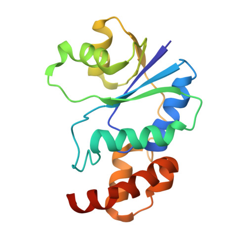
Structure of nicotinamide mononucleotide adenylyltransferase: a key enzyme in NAD(+) biosynthesis.

D'Angelo, I.Raffaelli, N.Dabusti, V.Lorenzi, T.Magni, G.Rizzi, M.

(2000) Structure 8: 993-1004

  • DOI: https://doi.org/10.1016/s0969-2126(00)00190-8
  • Primary Citation of Related Structures:  
    1F9A

  • PubMed Abstract: 

    Nicotinamide adenine dinucleotide (NAD(+)) is an essential cofactor involved in fundamental processes in cell metabolism. The enzyme nicotinamide mononucleotide adenylyltransferase (NMN AT) plays a key role in NAD(+) biosynthesis, catalysing the condensation of nicotinamide mononucleotide and ATP, and yielding NAD(+) and pyrophosphate. Given its vital role in cell life, the enzyme represents a possible target for the development of new antibacterial agents. The structure of NMN AT from Methanococcus jannaschii in complex with ATP has been solved by X-ray crystallography at 2.0 A resolution, using a combination of single isomorphous replacement and density modification techniques. The structure reveals a hexamer with 32 point group symmetry composed of alpha/beta topology subunits. The catalytic site is located in a deep cleft on the surface of each subunit, where one ATP molecule and one Mg(2+) are observed. A strictly conserved HXGH motif (in single-letter amino acid code) is involved in ATP binding and recognition. The structure of NMN AT closely resembles that of phosphopantetheine adenylyltransferase. Remarkably, in spite of the fact that the two enzymes share the same fold and hexameric assembly, a striking difference in their quaternary structure is observed. Moreover, on the basis of structural similarity including the HXGH motif, we identify NMN AT as a novel member of the newly proposed superfamily of nucleotidyltransferase alpha/beta phosphodiesterases. Our structural data suggest that the catalytic mechanism does not rely on the direct involvement of any protein residues and is likely to be carried out through optimal positioning of substrates and transition-state stabilisation, as is proposed for other members of the nucleotidyltransferase alpha/beta phosphodiesterase superfamily.


  • Organizational Affiliation

    Department of Genetics and Microbiology 'A. Buzzati Traverso', University of Pavia, via Ferrata 1, 27100, Pavia, Italy.


Macromolecules
Find similar proteins by:  (by identity cutoff)  |  3D Structure
Entity ID: 1
MoleculeChains Sequence LengthOrganismDetailsImage
HYPOTHETICAL PROTEIN MJ0541
A, B, C, D, E
A, B, C, D, E, F
168Methanocaldococcus jannaschiiMutation(s): 0 
UniProt
Find proteins for Q57961 (Methanocaldococcus jannaschii (strain ATCC 43067 / DSM 2661 / JAL-1 / JCM 10045 / NBRC 100440))
Explore Q57961 
Go to UniProtKB:  Q57961
Entity Groups  
Sequence Clusters30% Identity50% Identity70% Identity90% Identity95% Identity100% Identity
UniProt GroupQ57961
Sequence Annotations
Expand
  • Reference Sequence
Small Molecules
Ligands 2 Unique
IDChains Name / Formula / InChI Key2D Diagram3D Interactions
ATP
Query on ATP

Download Ideal Coordinates CCD File 
H [auth A]
J [auth B]
L [auth C]
N [auth D]
P [auth E]
H [auth A],
J [auth B],
L [auth C],
N [auth D],
P [auth E],
R [auth F]
ADENOSINE-5'-TRIPHOSPHATE
C10 H16 N5 O13 P3
ZKHQWZAMYRWXGA-KQYNXXCUSA-N
MG
Query on MG

Download Ideal Coordinates CCD File 
G [auth A]
I [auth B]
K [auth C]
M [auth D]
O [auth E]
G [auth A],
I [auth B],
K [auth C],
M [auth D],
O [auth E],
Q [auth F]
MAGNESIUM ION
Mg
JLVVSXFLKOJNIY-UHFFFAOYSA-N
Experimental Data & Validation

Experimental Data

  • Method: X-RAY DIFFRACTION
  • Resolution: 2.00 Å
  • R-Value Free: 0.264 
  • R-Value Work: 0.215 
  • R-Value Observed: 0.215 
  • Space Group: P 1 21 1
Unit Cell:
Length ( Å )Angle ( ˚ )
a = 78.768α = 90
b = 112.642β = 116.94
c = 79.869γ = 90
Software Package:
Software NamePurpose
SHELXSphasing
REFMACrefinement
MOSFLMdata reduction
CCP4data scaling

Structure Validation

View Full Validation Report


Entry History 

Deposition Data

Revision History  (Full details and data files)

  • Version 1.0: 2001-01-10
    Type: Initial release
  • Version 1.1: 2008-04-27
    Changes: Version format compliance
  • Version 1.2: 2011-07-13
    Changes: Version format compliance
  • Version 1.3: 2024-02-07
    Changes: Data collection, Database references, Derived calculations