1F12

L-3-HYDROXYACYL-COA DEHYDROGENASE COMPLEXED WITH 3-HYDROXYBUTYRYL-COA


Experimental Data Snapshot

  • Method: X-RAY DIFFRACTION
  • Resolution: 2.40 Å
  • R-Value Free: 0.265 
  • R-Value Work: 0.212 

wwPDB Validation   3D Report Full Report


Ligand Structure Quality Assessment 


This is version 1.3 of the entry. See complete history


Literature

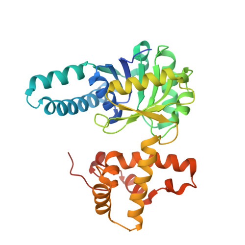
Sequestration of the active site by interdomain shifting. Crystallographic and spectroscopic evidence for distinct conformations of L-3-hydroxyacyl-CoA dehydrogenase.

Barycki, J.J.O'Brien, L.K.Strauss, A.W.Banaszak, L.J.

(2000) J Biol Chem 275: 27186-27196

  • DOI: https://doi.org/10.1074/jbc.M004669200
  • Primary Citation of Related Structures:  
    1F0Y, 1F12, 1F14, 1F17

  • PubMed Abstract: 
  • l-3-Hydroxyacyl-CoA dehydrogenase reversibly catalyzes the conversion of l-3-hydroxyacyl-CoA to 3-ketoacyl-CoA concomitant with the reduction of NAD(+) to NADH as part of the beta-oxidation spiral. In this report, crystal structures have been solved for the apoenzyme, binary complexes of the enzyme with reduced cofactor or 3-hydroxybutyryl-CoA substrate, and an abortive ternary complex of the enzyme with NAD(+) and acetoacetyl-CoA ...

    l-3-Hydroxyacyl-CoA dehydrogenase reversibly catalyzes the conversion of l-3-hydroxyacyl-CoA to 3-ketoacyl-CoA concomitant with the reduction of NAD(+) to NADH as part of the beta-oxidation spiral. In this report, crystal structures have been solved for the apoenzyme, binary complexes of the enzyme with reduced cofactor or 3-hydroxybutyryl-CoA substrate, and an abortive ternary complex of the enzyme with NAD(+) and acetoacetyl-CoA. The models illustrate positioning of cofactor and substrate within the active site of the enzyme. Comparison of these structures with the previous model of the enzyme-NAD(+) complex reveals that although significant shifting of the NAD(+)-binding domain relative to the C-terminal domain occurs in the ternary and substrate-bound complexes, there are few differences between the apoenzyme and cofactor-bound complexes. Analysis of these models clarifies the role of key amino acids implicated in catalysis and highlights additional critical residues. Furthermore, a novel charge transfer complex has been identified in the course of abortive ternary complex formation, and its characterization provides additional insight into aspects of the catalytic mechanism of l-3-hydroxyacyl-CoA dehydrogenase.


    Related Citations: 
    • Biochemical Characterization and Crystal Structure Determination of Human Heart Short Chain L-3-hydroxyacyl-CoA Dehydrogenase Provide Insights into Catalytic Mechanism
      Barycki, J.J., O'Brien, L.K., Bratt, J.M., Zhang, R., Sanishvili, R., Strauss, A.W., Banaszak, L.J.
      (1999) Biochemistry 38: 5786

    Organizational Affiliation

    Department of Biochemistry, Molecular Biology, and Biophysics, University of Minnesota, Minneapolis, Minnesota 55455, USA.


Macromolecules
Find similar proteins by:  (by identity cutoff)  |  3D Structure
Entity ID: 1
MoleculeChainsSequence LengthOrganismDetailsImage
L-3-HYDROXYACYL-COA DEHYDROGENASE
A, B
310Homo sapiensMutation(s): 1 
Gene Names: HADHHADHAD1HADHSCSCHAD
EC: 1.1.1.35
UniProt & NIH Common Fund Data Resources
Find proteins for Q16836 (Homo sapiens)
Explore Q16836 
Go to UniProtKB:  Q16836
PHAROS:  Q16836
Entity Groups  
Sequence Clusters30% Identity50% Identity70% Identity90% Identity95% Identity100% Identity
UniProt GroupQ16836
Protein Feature View
Expand
  • Reference Sequence
Small Molecules
Ligands 1 Unique
IDChainsName / Formula / InChI Key2D Diagram3D Interactions
3HC
Query on 3HC

Download Ideal Coordinates CCD File 
C [auth A]3-HYDROXYBUTANOYL-COENZYME A
C25 H42 N7 O18 P3 S
QHHKKMYHDBRONY-VKBDFPRVSA-N
 Ligand Interaction
Experimental Data & Validation

Experimental Data

  • Method: X-RAY DIFFRACTION
  • Resolution: 2.40 Å
  • R-Value Free: 0.265 
  • R-Value Work: 0.212 
  • Space Group: P 21 21 21
Unit Cell:
Length ( Å )Angle ( ˚ )
a = 50.04α = 90
b = 88.1β = 90
c = 167.22γ = 90
Software Package:
Software NamePurpose
CNSrefinement
DENZOdata reduction
SCALEPACKdata scaling
CNSphasing

Structure Validation

View Full Validation Report


Ligand Structure Quality Assessment 


Entry History 

Deposition Data

Revision History  (Full details and data files)

  • Version 1.0: 2000-09-27
    Type: Initial release
  • Version 1.1: 2008-04-27
    Changes: Version format compliance
  • Version 1.2: 2011-07-13
    Changes: Version format compliance
  • Version 1.3: 2021-11-03
    Changes: Database references, Derived calculations