Structural analysis of the transcriptional activation on Fis: crystal structures of six Fis mutants with different activation properties.
Cheng, Y.S., Yang, W.Z., Johnson, R.C., Yuan, H.S.(2000) J Mol Biol 302: 1139-1151
- PubMed: 11183780 Search on PubMed
- DOI: https://doi.org/10.1006/jmbi.2000.4123
- Primary Citation Related Structures:
1ETK, 1ETO, 1ETQ, 1ETV, 1ETW, 1ETX, 1ETY - PubMed Abstract:
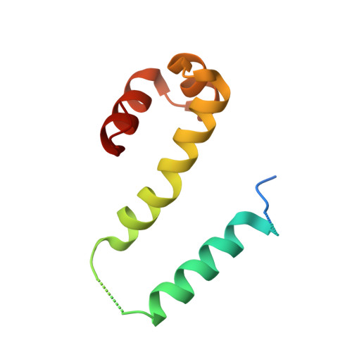
The Fis protein regulates gene expression in Escherichia coli by activating or repressing transcription of a variety of genes. Fis can activate transcription when bound to DNA upstream of the RNA-polymerase-binding site, such as in the rrnB P1 promoter, or when bound to a site overlapping the -35 RNA polymerase binding site, such as in the proP P2 promoter. It has been suggested that transcriptional activation in both promoters results from interactions between specific amino acids within a turn connecting the B and C helices (the BC turn) in Fis and the C-terminal domain of the alpha-subunit of RNA polymerase (alphaCTD of RNAP). Here, crystal structures of six Fis BC turn mutants with different transcriptional activation properties, Q68A, R71Y, R71L, G72A, G72D and Q74A, were determined at 1.9 to 2.8 A resolution. Two of these mutants, R71Y and R71L, crystallized in unit cells which are different from that of wild-type Fis, and the structure of R71L offers the most complete Fis model to date in that the extended structure of the N-terminal region is revealed. The BC turn in all of these mutant structures remains in a nearly identical gamma gamma beta-turn conformation as present in wild-type Fis. Analyses of the molecular surfaces of the transactivation region of the mutants suggest that several residues in or near the BC turn, including Gln68, Arg71, Gly72 and Gln74, form a ridge that could contact the alphaCTD of RNAP on one side. The structures and biochemical properties of the mutants suggest that Arg71 is the most critical residue for contacting RNAP within this ridge and that the glycine at position 72 helps to stabilize the structure.
- Graduate Institute of Life Science, National Defense Medical Center, Taipei, Taiwan, Republic of China.
Organizational Affiliation: