1EEX

CRYSTAL STRUCTURE OF THE DIOL DEHYDRATASE-ADENINYLPENTYLCOBALAMIN COMPLEX FROM KLEBSIELLA OXYTOCA


Experimental Data Snapshot

  • Method: X-RAY DIFFRACTION
  • Resolution: 1.70 Å
  • R-Value Free: 0.226 
  • R-Value Work: 0.161 
  • R-Value Observed: 0.164 

wwPDB Validation   3D Report Full Report


Ligand Structure Quality Assessment 


This is version 1.5 of the entry. See complete history


Literature

How a protein generates a catalytic radical from coenzyme B(12): X-ray structure of a diol-dehydratase-adeninylpentylcobalamin complex.

Masuda, J.Shibata, N.Morimoto, Y.Toraya, T.Yasuoka, N.

(2000) Structure 8: 775-788

  • DOI: https://doi.org/10.1016/s0969-2126(00)00164-7
  • Primary Citation of Related Structures:  
    1EEX, 1EGM, 1EGV

  • PubMed Abstract: 

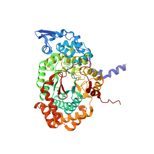
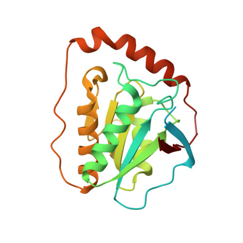
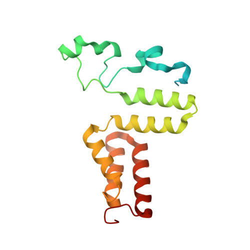
    Adenosylcobalamin (coenzyme B(12)) serves as a cofactor for enzymatic radical reactions. The adenosyl radical, a catalytic radical in these reactions, is formed by homolysis of the cobalt-carbon bond of the coenzyme, although the mechanism of cleavage of its organometallic bond remains unsolved. We determined the three-dimensional structures of diol dehydratase complexed with adeninylpentylcobalamin and with cyanocobalamin at 1.7 A and 1.9 A resolution, respectively, at cryogenic temperatures. In the adeninylpentylcobalamin complex, the adenine ring is bound parallel to the corrin ring as in the free form and methylmalonyl-CoA-mutase-bound coenzyme, but with the other side facing pyrrole ring C. All of its nitrogen atoms except for N(9) are hydrogen-bonded to mainchain amide oxygen and amide nitrogen atoms, a sidechain hydroxyl group, and a water molecule. As compared with the cyanocobalamin complex, the sidechain of Seralpha224 rotates by 120 degrees to hydrogen bond with N(3) of the adenine ring. The structure of the adenine-ring-binding site provides a molecular basis for the strict specificity of diol dehydratase for the coenzyme adenosyl group. The superimposition of the structure of the free coenzyme on that of enzyme-bound adeninylpentylcobalamin demonstrated that the tight enzyme-coenzyme interactions at both the cobalamin moiety and adenine ring of the adenosyl group would inevitably lead to cleavage of the cobalt-carbon bond. Rotation of the ribose moiety around the glycosidic linkage makes the 5'-carbon radical accessible to the hydrogen atom of the substrate to be abstracted.


  • Organizational Affiliation

    Department of Life Science, Himeji Institute of Technology, Kamigori, Akogun, 678-1297, Japan.


Macromolecules
Find similar proteins by:  (by identity cutoff)  |  3D Structure
Entity ID: 1
MoleculeChains Sequence LengthOrganismDetailsImage
PROPANEDIOL DEHYDRATASEA,
D [auth L]
554Klebsiella oxytocaMutation(s): 0 
EC: 4.2.1.28
UniProt
Find proteins for Q59470 (Klebsiella oxytoca)
Explore Q59470 
Go to UniProtKB:  Q59470
Entity Groups  
Sequence Clusters30% Identity50% Identity70% Identity90% Identity95% Identity100% Identity
UniProt GroupQ59470
Sequence Annotations
Expand
  • Reference Sequence
Find similar proteins by:  (by identity cutoff)  |  3D Structure
Entity ID: 2
MoleculeChains Sequence LengthOrganismDetailsImage
PROPANEDIOL DEHYDRATASE
B, E
224Klebsiella oxytocaMutation(s): 0 
EC: 4.2.1.28
UniProt
Find proteins for Q59471 (Klebsiella oxytoca)
Explore Q59471 
Go to UniProtKB:  Q59471
Entity Groups  
Sequence Clusters30% Identity50% Identity70% Identity90% Identity95% Identity100% Identity
UniProt GroupQ59471
Sequence Annotations
Expand
  • Reference Sequence
Find similar proteins by:  (by identity cutoff)  |  3D Structure
Entity ID: 3
MoleculeChains Sequence LengthOrganismDetailsImage
PROPANEDIOL DEHYDRATASEC [auth G],
F [auth M]
173Klebsiella oxytocaMutation(s): 0 
EC: 4.2.1.28
UniProt
Find proteins for Q59472 (Klebsiella oxytoca)
Explore Q59472 
Go to UniProtKB:  Q59472
Entity Groups  
Sequence Clusters30% Identity50% Identity70% Identity90% Identity95% Identity100% Identity
UniProt GroupQ59472
Sequence Annotations
Expand
  • Reference Sequence
Experimental Data & Validation

Experimental Data

  • Method: X-RAY DIFFRACTION
  • Resolution: 1.70 Å
  • R-Value Free: 0.226 
  • R-Value Work: 0.161 
  • R-Value Observed: 0.164 
  • Space Group: P 21 21 21
Unit Cell:
Length ( Å )Angle ( ˚ )
a = 73.38α = 90
b = 121.4β = 90
c = 207.6γ = 90
Software Package:
Software NamePurpose
SHELXL-97refinement
CNSrefinement
MOSFLMdata reduction
CCP4data scaling
CNSphasing

Structure Validation

View Full Validation Report


Ligand Structure Quality Assessment 


Entry History 

Deposition Data

Revision History  (Full details and data files)

  • Version 1.0: 2001-02-07
    Type: Initial release
  • Version 1.1: 2008-04-27
    Changes: Version format compliance
  • Version 1.2: 2011-07-13
    Changes: Version format compliance
  • Version 1.3: 2019-07-24
    Changes: Data collection, Refinement description
  • Version 1.4: 2024-02-07
    Changes: Data collection, Database references, Derived calculations
  • Version 1.5: 2024-03-13
    Changes: Source and taxonomy, Structure summary