1EBD

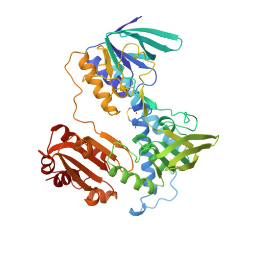
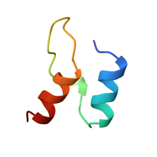
DIHYDROLIPOAMIDE DEHYDROGENASE COMPLEXED WITH THE BINDING DOMAIN OF THE DIHYDROLIPOAMIDE ACETYLASE


Experimental Data Snapshot

  • Method: X-RAY DIFFRACTION
  • Resolution: 2.60 Å
  • R-Value Work: 0.215 
  • R-Value Observed: 0.215 

wwPDB Validation   3D Report Full Report


This is version 1.2 of the entry. See complete history


Literature

Protein-protein interactions in the pyruvate dehydrogenase multienzyme complex: dihydrolipoamide dehydrogenase complexed with the binding domain of dihydrolipoamide acetyltransferase.

Mande, S.S.Sarfaty, S.Allen, M.D.Perham, R.N.Hol, W.G.

(1996) Structure 4: 277-286

  • DOI: https://doi.org/10.1016/s0969-2126(96)00032-9
  • Primary Citation of Related Structures:  
    1EBD

  • PubMed Abstract: 

    The ubiquitous pyruvate dehydrogenase multienzyme complex is built around an octahedral or icosahedral core of dihydrolipoamide acetyltransferase (E2) chains, to which multiple copies of pyruvate decarboxylase (E1) and dihydrolipoamide dehydrogenase (E3) bind tightly but non-covalently. E2 is a flexible multidomain protein that mediates interactions with E1 and E3 through a remarkably small binding domain (E2BD). In the Bacillus stearothermophilus complex, the E2 core is an icosahedral assembly of 60 E2 chains. The crystal structure of the E3 dimer (101 kDa) complexed with E2BD (4 kDa) has been solved to 2.6 A resolution. Interactions between E3 and E2BD are dominated by an electrostatic zipper formed by Arg135 and Arg139 in the N-terminal helix of E2BD and Asp344 and Glu431 of one of the monomers of E3. E2BD interacts with both E3 monomers, but the binding site is located close to the twofold axis. Thus, in agreement with earlier biochemical results, it is impossible for two molecules of E2BD to bind simultaneously to one E3 dimer. Combining this new structure for the E3-E2BD complex with previously determined structures of the E2 catalytic domain and the E2 lipoyl domain creates a model of the E2 core showing how the lipoyl domain can move between the active sites of E2 and E3 in the multienzyme complex.


  • Organizational Affiliation

    Department of Biological Structure, Biomolecular Structure Center, University of Washington, Box 357742, Seattle, WA 98195, USA.


Macromolecules
Find similar proteins by:  (by identity cutoff)  |  3D Structure
Entity ID: 1
MoleculeChains Sequence LengthOrganismDetailsImage
DIHYDROLIPOAMIDE DEHYDROGENASE
A, B
455Geobacillus stearothermophilusMutation(s): 0 
EC: 1.8.1.4
UniProt
Find proteins for P11959 (Geobacillus stearothermophilus)
Explore P11959 
Go to UniProtKB:  P11959
Entity Groups  
Sequence Clusters30% Identity50% Identity70% Identity90% Identity95% Identity100% Identity
UniProt GroupP11959
Sequence Annotations
Expand
  • Reference Sequence
Find similar proteins by:  (by identity cutoff)  |  3D Structure
Entity ID: 2
MoleculeChains Sequence LengthOrganismDetailsImage
DIHYDROLIPOAMIDE ACETYLTRANSFERASE41Geobacillus stearothermophilusMutation(s): 0 
UniProt
Find proteins for P11961 (Geobacillus stearothermophilus)
Explore P11961 
Go to UniProtKB:  P11961
Entity Groups  
Sequence Clusters30% Identity50% Identity70% Identity90% Identity95% Identity100% Identity
UniProt GroupP11961
Sequence Annotations
Expand
  • Reference Sequence
Small Molecules
Ligands 1 Unique
IDChains Name / Formula / InChI Key2D Diagram3D Interactions
FAD
Query on FAD

Download Ideal Coordinates CCD File 
D [auth A],
E [auth B]
FLAVIN-ADENINE DINUCLEOTIDE
C27 H33 N9 O15 P2
VWWQXMAJTJZDQX-UYBVJOGSSA-N
Experimental Data & Validation

Experimental Data

  • Method: X-RAY DIFFRACTION
  • Resolution: 2.60 Å
  • R-Value Work: 0.215 
  • R-Value Observed: 0.215 
  • Space Group: P 31 2 1
Unit Cell:
Length ( Å )Angle ( ˚ )
a = 106.6α = 90
b = 106.6β = 90
c = 204.3γ = 120
Software Package:
Software NamePurpose
DENZOdata reduction
SCALEPACKdata scaling
X-PLORmodel building
X-PLORrefinement
X-PLORphasing

Structure Validation

View Full Validation Report


Entry History 

Deposition Data

Revision History  (Full details and data files)

  • Version 1.0: 1996-07-11
    Type: Initial release
  • Version 1.1: 2008-03-24
    Changes: Version format compliance
  • Version 1.2: 2011-07-13
    Changes: Version format compliance