1E9L

The crystal structure of novel mammalian lectin Ym1 suggests a saccharide binding site


Experimental Data Snapshot

  • Method: X-RAY DIFFRACTION
  • Resolution: 2.50 Å
  • R-Value Free: 0.272 
  • R-Value Work: 0.198 
  • R-Value Observed: 0.198 

wwPDB Validation   3D Report Full Report


This is version 2.2 of the entry. See complete history


Literature

The Crystal Structure of a Novel Mammalian Lectin, Ym1, Suggests a Saccharide Binding Site

Sun, Y.J.Chang, N.C.Hung, S.I.Chang, A.C.Chou, C.C.Hsiao, C.D.

(2001) J Biol Chem 276: 17507

  • DOI: https://doi.org/10.1074/jbc.M010416200
  • Primary Citation of Related Structures:  
    1E9L

  • PubMed Abstract: 

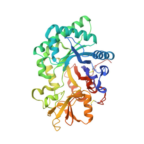
    Ym1, a secretory protein synthesized by activated murine peritoneal macrophages, is a novel mammalian lectin with a binding specificity to GlcN. Lectins are responsible for carbohydrate recognition and for mediating cell-cell and cell-extracellular matrix interactions in microbes, plants, and animals. Glycosaminoglycan heparin/heparan sulfate binding ability was also detected in Ym1. We report here the three-dimensional structure of Ym1 at 2.5-A resolution by x-ray crystallography. The crystal structure of Ym1 consists of two globular domains, a beta/alpha triose-phosphate isomerase barrel domain and a small alpha + beta folding domain. A notable electron density of sugar is detected in the Ym1 crystal structure. The saccharide is located inside the triose-phosphate isomerase domain at the COOH terminal end of the beta-strands. Both hydrophilic and hydrophobic interactions are noted in the sugar-binding site in Ym1. Despite the fact that Ym1 is not a chitinase, structurally, Ym1 shares significant homology with chitinase A of Serratia marcescens. Ym1 and chitinase A have a similar carbohydrate binding cleft. This study provides new structure information, which will lead to better understanding of the biological significance of Ym1 and its putative gene members.


  • Organizational Affiliation

    Institute of Molecular Biology, Academia Sinica, Taipei, Taiwan 115, Republic of China.


Macromolecules
Find similar proteins by:  (by identity cutoff)  |  3D Structure
Entity ID: 1
MoleculeChains Sequence LengthOrganismDetailsImage
YM1 SECRETORY PROTEIN377Mus musculusMutation(s): 0 
UniProt
Find proteins for O35744 (Mus musculus)
Explore O35744 
Go to UniProtKB:  O35744
Entity Groups  
Sequence Clusters30% Identity50% Identity70% Identity90% Identity95% Identity100% Identity
UniProt GroupO35744
Sequence Annotations
Expand
  • Reference Sequence
Small Molecules
Ligands 1 Unique
IDChains Name / Formula / InChI Key2D Diagram3D Interactions
GCS
Query on GCS

Download Ideal Coordinates CCD File 
B [auth A]2-amino-2-deoxy-beta-D-glucopyranose
C6 H13 N O5
MSWZFWKMSRAUBD-QZABAPFNSA-N
Experimental Data & Validation

Experimental Data

  • Method: X-RAY DIFFRACTION
  • Resolution: 2.50 Å
  • R-Value Free: 0.272 
  • R-Value Work: 0.198 
  • R-Value Observed: 0.198 
  • Space Group: P 1 21 1
Unit Cell:
Length ( Å )Angle ( ˚ )
a = 51.34α = 90
b = 60.66β = 94.61
c = 60.76γ = 90
Software Package:
Software NamePurpose
X-PLORrefinement
DENZOdata reduction
SCALEPACKdata scaling
CCP4phasing

Structure Validation

View Full Validation Report


Entry History 

Deposition Data

Revision History  (Full details and data files)

  • Version 1.0: 2001-03-01
    Type: Initial release
  • Version 1.1: 2011-05-08
    Changes: Version format compliance
  • Version 1.2: 2011-07-13
    Changes: Version format compliance
  • Version 2.0: 2019-05-22
    Changes: Atomic model, Data collection, Derived calculations, Other, Refinement description
  • Version 2.1: 2020-07-01
    Changes: Advisory, Data collection, Derived calculations, Other
  • Version 2.2: 2020-07-29
    Type: Remediation
    Reason: Carbohydrate remediation
    Changes: Data collection, Derived calculations, Structure summary