1E0F

Crystal structure of the human alpha-thrombin-haemadin complex: an exosite II-binding inhibitor


Experimental Data Snapshot

  • Method: X-RAY DIFFRACTION
  • Resolution: 3.10 Å
  • R-Value Free: 0.255 
  • R-Value Work: 0.208 
  • R-Value Observed: 0.208 

wwPDB Validation   3D Report Full Report


This is version 1.3 of the entry. See complete history


Literature

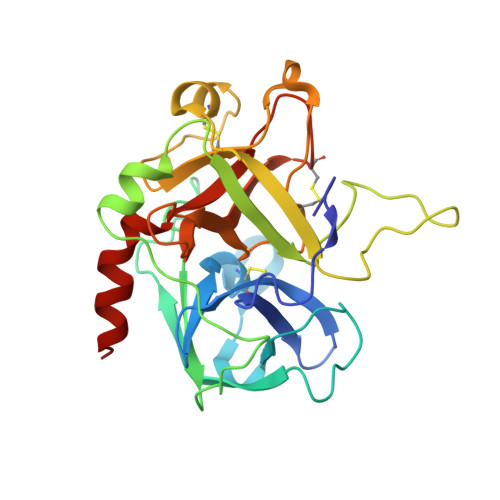
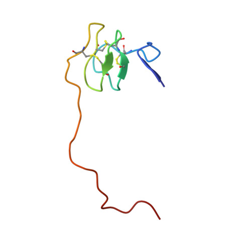
Crystal Structure of the Human Alpha-Thrombin-Haemadin Complex: An Exosite II-Binding Inhibitor

Richardson, J.L.Kroeger, B.Hoeffken, W.Sadler, J.E.Pereira, P.Huber, R.Bode, W.Fuentes-Prior, P.

(2000) EMBO J 19: 5650

  • DOI: 10.1093/emboj/19.21.5650
  • Primary Citation of Related Structures:  
    1E0F

  • PubMed Abstract: 
  • The serine proteinase alpha-thrombin plays a pivotal role in the regulation of blood fluidity, and therefore constitutes a primary target in the treatment of various haemostatic disorders. Haemadin is a slow tight- binding thrombin inhibitor from the land-living leech Haemadipsa sylvestris ...

    The serine proteinase alpha-thrombin plays a pivotal role in the regulation of blood fluidity, and therefore constitutes a primary target in the treatment of various haemostatic disorders. Haemadin is a slow tight- binding thrombin inhibitor from the land-living leech Haemadipsa sylvestris. Here we present the 3.1 A crystal structure of the human alpha-thrombin- haemadin complex. The N-terminal segment of haemadin binds to the active site of thrombin, forming a parallel beta-strand with residues Ser214-Gly216 of the proteinase. This mode of binding is similar to that observed in another leech-derived inhibitor, hirudin. In contrast to hirudin, however, the markedly acidic C-terminal peptide of haemadin does not bind the fibrinogen-recognition exosite, but interacts with the heparin-binding exosite of thrombin. Thus, haemadin binds to thrombin according to a novel mechanism, despite an overall structural similarity with hirudin. Haemadin inhibits both free and thrombomodulin-bound alpha-thrombin, but not intermediate activation forms such as meizothrombin. This specific anticoagulant ability of haemadin makes it an ideal candidate for an antithrombotic agent, as well as a starting point for the design of novel antithrombotics.


    Related Citations: 
    • Isolation, Sequence Analysis, and Cloning of Haemadin an Anticoagulant Peptide from the Indian Leech
      Strube, K.-H., Kroeger, B., Bialojan, S., Otte, M., Dodt, J.
      (1993) J Biol Chem 268: 8590
    • The Refined 1.9 Angstrom X-Ray Crystal Structure of D-Phe-Pro-Arg-Chloromethylketone Inhibited Human Alpha Thrombin: Structure Analysis, Overall Structure, Electrostatic Properties, Detailed Active Site Geometry and Structure Function Relatioships
      Bode, W., Turk, D., Karshikov, A.
      (1992) Protein Sci 1: 426

    Organizational Affiliation

    Max-Planck-Institut für Biochemie, D-82152 Martinsried, Department of Biotechnology, BASF Aktiengesellschaft, D-67056 Ludwigshafen, Germany. richards@biochem.mpg.de


Macromolecules
Find similar proteins by:  (by identity cutoff)  |  3D Structure
Entity ID: 1
MoleculeChainsSequence LengthOrganismDetailsImage
THROMBINA, B, C36Homo sapiensMutation(s): 0 
Gene Names: F2
EC: 3.4.21.5
UniProt & NIH Common Fund Data Resources
Find proteins for P00734 (Homo sapiens)
Explore P00734 
Go to UniProtKB:  P00734
PHAROS:  P00734
Entity Groups  
Sequence Clusters30% Identity50% Identity70% Identity90% Identity95% Identity100% Identity
UniProt GroupP00734
Protein Feature View
Expand
  • Reference Sequence
Find similar proteins by:  (by identity cutoff)  |  3D Structure
Entity ID: 2
MoleculeChainsSequence LengthOrganismDetailsImage
THROMBIND, E, F259Homo sapiensMutation(s): 0 
Gene Names: F2
EC: 3.4.21.5
UniProt & NIH Common Fund Data Resources
Find proteins for P00734 (Homo sapiens)
Explore P00734 
Go to UniProtKB:  P00734
PHAROS:  P00734
Entity Groups  
Sequence Clusters30% Identity50% Identity70% Identity90% Identity95% Identity100% Identity
UniProt GroupP00734
Protein Feature View
Expand
  • Reference Sequence
Find similar proteins by:  (by identity cutoff)  |  3D Structure
Entity ID: 3
MoleculeChainsSequence LengthOrganismDetailsImage
HAEMADING [auth I],
H [auth J],
I [auth K]
57Haemadipsa sylvestrisMutation(s): 0 
UniProt
Find proteins for Q25163 (Haemadipsa sylvestris)
Explore Q25163 
Go to UniProtKB:  Q25163
Entity Groups  
Sequence Clusters30% Identity50% Identity70% Identity90% Identity95% Identity100% Identity
UniProt GroupQ25163
Protein Feature View
Expand
  • Reference Sequence
Experimental Data & Validation

Experimental Data

  • Method: X-RAY DIFFRACTION
  • Resolution: 3.10 Å
  • R-Value Free: 0.255 
  • R-Value Work: 0.208 
  • R-Value Observed: 0.208 
  • Space Group: P 1 2 1
Unit Cell:
Length ( Å )Angle ( ˚ )
a = 121.67α = 90
b = 50.57β = 114.76
c = 129.74γ = 90
Software Package:
Software NamePurpose
X-PLORrefinement
MOSFLMdata reduction
Agrovatadata scaling
ROTAVATAdata scaling
AMoREphasing

Structure Validation

View Full Validation Report


Entry History 

Deposition Data

Revision History  (Full details and data files)

  • Version 1.0: 2000-11-03
    Type: Initial release
  • Version 1.1: 2012-11-21
    Changes: Database references, Derived calculations, Other, Source and taxonomy, Structure summary
  • Version 1.2: 2017-07-05
    Changes: Data collection
  • Version 1.3: 2019-05-08
    Changes: Data collection, Experimental preparation