Structure of the C-Terminal Sterile Alpha-Motif (Sam) Domain of Human P73 Alpha
Wang, W.K., Bycroft, M., Foster, N.W., Buckle, A.M., Fersht, A.R., Chen, Y.W.(2001) Acta Crystallogr D Biol Crystallogr 57: 545
- PubMed: 11264583 Search on PubMed
- DOI: https://doi.org/10.1107/s0907444901002529
- Primary Citation Related Structures:
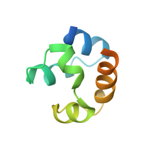
1DXS - PubMed Abstract:
p73 is a homologue of the tumour suppressor p53 and contains all three functional domains of p53. The alpha-splice variant of p73 (p73 alpha) contains near its C-terminus an additional structural domain known as the sterile alpha-motif (SAM) that is probably responsible for regulating p53-like functions of p73. Here, the 2.54 A resolution crystal structure of this protein domain is reported. The crystal structure and the published solution structure have the same five-helix bundle fold that is characteristic of all SAM-domain structures, with an overall r.m.s.d. of 1.5 A for main-chain atoms. The hydrophobic core residues are well conserved, yet some large local differences are observed. The crystal structure reveals a dimeric organization, with the interface residues forming a mini four-helix bundle. However, analysis of solvation free energies and the surface area buried upon dimer formation indicated that this arrangement is more likely to be an effect of crystal packing rather than reflecting a physiological state. This is consistent with the solution structure being a monomer. The p73 alpha SAM domain also contains several interesting structural features: a Cys-X-X-Cys motif, a 3(10)-helix and a loop that have elevated B factors, and short tight inter-helical loops including two beta-turns; these elements are probably important in the normal function of this domain.
- Centre for Protein Engineering and Cambridge University Chemical Laboratory, MRC Centre, Hills Road, Cambridge CB2 2QH, England.
Organizational Affiliation: