A structural requirement for activation of skeletal ryanodine receptors by peptides of the dihydropyridine receptor II-III loop.
Casarotto, M.G., Gibson, F., Pace, S.M., Curtis, S.M., Mulcair, M., Dulhunty, A.F.(2000) J Biological Chem 275: 11631-11637
- PubMed: 10766780 Search on PubMed
- DOI: https://doi.org/10.1074/jbc.275.16.11631
- Primary Citation Related Structures:
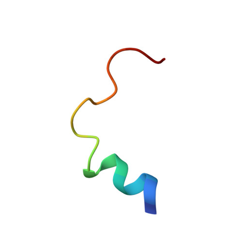
1DU1 - PubMed Abstract:
The solution structures of three related peptides (A1, A2, and A9) corresponding to the Thr(671)-Leu(690) region of the skeletal muscle dihydropyridine receptor II-III loop have been investigated using nuclear magnetic resonance spectroscopy. Peptide A1, the native sequence, is less effective in activating ryanodine receptor calcium release channels than A2 (Ser(687) to Ala substitution). Peptide A9, Arg(681)-Ser(687), does not activate ryanodine receptors. A1 and A2 are helical from their N terminus to Lys(685) but are generally unstructured from Lys(685) to the C terminus. The basic residues Arg(681)-Lys(685), essential for A1 activation of ryanodine receptors, are located at the C-terminal end of the alpha-helix. Peptide A9 was found to be unstructured. Differences between A1 and A2 were observed in the C-terminal end of the helix (residues 681-685), which was less ordered in A1, and in the C-terminal region of the peptide, which exhibited greater flexibility in A1. Predicted low energy models suggest that an electrostatic interaction between the hydroxyl oxygen of Ser(687) and the guanidino moiety of Arg(683) is lost with the Ser(687)Ala substitution. The results show that the more structured peptides are more effective in activating ryanodine receptors.
- Division of Biochemistry and Molecular Biology, John Curtin School of Medical Research, Australian National University, Canberra, ACT 2601, Australia. macro.casarotto@anu.edu.au
Organizational Affiliation: