1D0L

THE ESCHERICHIA COLI LYTIC TRANSGLYCOSYLASE SLT35 IN COMPLEX WITH BULGECIN A


Experimental Data Snapshot

  • Method: X-RAY DIFFRACTION
  • Resolution: 1.97 Å
  • R-Value Free: 0.200 
  • R-Value Work: 0.168 
  • R-Value Observed: 0.170 

wwPDB Validation   3D Report Full Report


Ligand Structure Quality Assessment 


This is version 1.4 of the entry. See complete history


Literature

Crystallographic studies of the interactions of Escherichia coli lytic transglycosylase Slt35 with peptidoglycan.

van Asselt, E.J.Kalk, K.H.Dijkstra, B.W.

(2000) Biochemistry 39: 1924-1934

  • DOI: https://doi.org/10.1021/bi992161p
  • Primary Citation of Related Structures:  
    1D0K, 1D0L, 1D0M

  • PubMed Abstract: 

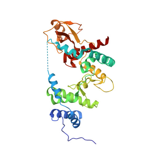
    Lytic transglycosylases catalyze the cleavage of the beta-1, 4-glycosidic bond between N-acetylmuramic acid (MurNAc) and N-acetylglucosamine (GlcNAc) in peptidoglycan with concomitant formation of a 1,6-anhydro bond in the MurNAc residue. To understand the reaction mechanism of Escherichia coli lytic transglycosylase Slt35, three crystal structures have been determined of Slt35 in complex with two different peptidoglycan fragments and with the lytic transglycosylase inhibitor bulgecin A. The complexes define four sugar-binding subsites (-2, -1, +1, and +2) and two peptide-binding sites in a large cleft close to Glu162. The Glu162 side chain is between the -1 and +1 sugar-binding sites, in agreement with a function as catalytic acid/base. The complexes suggest additional contributions to catalysis from Ser216 and Asn339, residues which are conserved among the MltB/Slt35 lytic transglycosylases.


  • Organizational Affiliation

    BIOSON Research Institute, Laboratory of Biophysical Chemistry, University of Groningen, Nijenborgh 4, 9747 AG Groningen, The Netherlands.


Macromolecules
Find similar proteins by:  (by identity cutoff)  |  3D Structure
Entity ID: 1
MoleculeChains Sequence LengthOrganismDetailsImage
35KD SOLUBLE LYTIC TRANSGLYCOSYLASE322Escherichia coliMutation(s): 2 
UniProt
Find proteins for P41052 (Escherichia coli (strain K12))
Explore P41052 
Go to UniProtKB:  P41052
Entity Groups  
Sequence Clusters30% Identity50% Identity70% Identity90% Identity95% Identity100% Identity
UniProt GroupP41052
Sequence Annotations
Expand
  • Reference Sequence
Small Molecules
Ligands 2 Unique
IDChains Name / Formula / InChI Key2D Diagram3D Interactions
BLG
Query on BLG

Download Ideal Coordinates CCD File 
C [auth A]4-O-(4-O-SULFONYL-N-ACETYLGLUCOSAMININYL)-5-METHYLHYDROXY-L-PROLINE-TAURINE
C16 H30 N3 O14 S2
RPNZWZDLNYCCIG-HMMVDTEZSA-O
CA
Query on CA

Download Ideal Coordinates CCD File 
B [auth A]CALCIUM ION
Ca
BHPQYMZQTOCNFJ-UHFFFAOYSA-N
Experimental Data & Validation

Experimental Data

  • Method: X-RAY DIFFRACTION
  • Resolution: 1.97 Å
  • R-Value Free: 0.200 
  • R-Value Work: 0.168 
  • R-Value Observed: 0.170 
  • Space Group: P 21 21 21
Unit Cell:
Length ( Å )Angle ( ˚ )
a = 58.395α = 90
b = 67.785β = 90
c = 97.735γ = 90
Software Package:
Software NamePurpose
X-PLORmodel building
X-PLORrefinement
DENZOdata reduction
SCALEPACKdata scaling
X-PLORphasing

Structure Validation

View Full Validation Report


Ligand Structure Quality Assessment 


Entry History 

Deposition Data

Revision History  (Full details and data files)

  • Version 1.0: 2000-03-06
    Type: Initial release
  • Version 1.1: 2008-04-27
    Changes: Version format compliance
  • Version 1.2: 2011-07-13
    Changes: Version format compliance
  • Version 1.3: 2021-11-03
    Changes: Data collection, Database references, Derived calculations
  • Version 1.4: 2024-02-07
    Changes: Data collection