1BJ3

CRYSTAL STRUCTURE OF COAGULATION FACTOR IX-BINDING PROTEIN (IX-BP) FROM VENOM OF HABU SNAKE WITH A HETERODIMER OF C-TYPE LECTIN DOMAINS


Experimental Data Snapshot

  • Method: X-RAY DIFFRACTION
  • Resolution: 2.60 Å
  • R-Value Free: 0.271 
  • R-Value Work: 0.182 
  • R-Value Observed: 0.182 

wwPDB Validation   3D Report Full Report


This is version 1.3 of the entry. See complete history


Literature

Crystal structure of coagulation factor IX-binding protein from habu snake venom at 2.6 A: implication of central loop swapping based on deletion in the linker region.

Mizuno, H.Fujimoto, Z.Koizumi, M.Kano, H.Atoda, H.Morita, T.

(1999) J Mol Biol 289: 103-112

  • DOI: https://doi.org/10.1006/jmbi.1999.2756
  • Primary Citation of Related Structures:  
    1BJ3

  • PubMed Abstract: 

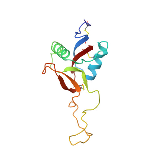
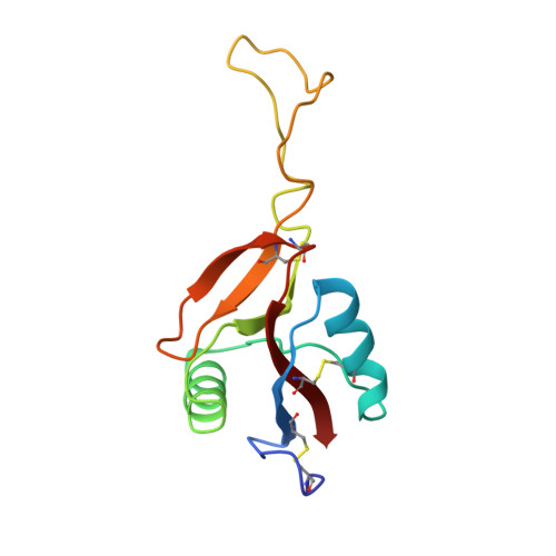
    Coagulation factor IX-binding protein (IX-bp) isolated from the venom of the habu snake (Trimeresurus flavoviridis) is a disulfide-linked heterodimer consisting of homologous subunits A and B. The structure of IX-bp has been solved by X-ray crystallography at 2.6 A resolution to a crystallographic R -value of 0.181. The main-chain fold of each subunit is homologous to the carbohydrate-recognition domain of C-type lectins (C-type CRDs) except for the extended central loop. The structure is almost identical with that of factors IX and X-binding protein (IX/X-bp) as expected from the high level of amino acid sequence homology. The functional difference in ligand recognition from IX/X-bp must reside in the amino acid differences. A continuity of different amino acid residues located from the C-terminal of the second alpha-helix to the following loop forms the local conformational difference in this region between the two proteins. This loop participates in the formation of the concave surface between the two subunits, the putative binding site for the Gla-domain (gamma-carboxyglutamic acid-containing domain) of the coagulation factors. Another difference between the two proteins is in the relative disposition of subunits A and B. When the B subunits are superimposed, about a 6 degrees rotation is required for the superposition of the A subunits. A calcium ion links the second alpha-helix region to the C-terminal tail in each subunit and helps to stabilize the structure for Gla-domain binding. The interface created by the central loop swapping in the dimer IX-bp is almost identical with that seen within the monomeric C-type CRDs. This dimer forms as the result of the amino acid deletion in the linker region of the central loop of the original C-type lectins. Such a dimerization disrupts the lectin active site and creates a Gla-domain binding site, imparting functional diversity.


  • Organizational Affiliation

    National Institute of Agrobiological Resources, Tsukuba Science City, Ibaraki, 305-8602, Japan. mizuno@abr.affrc.go.jp


Macromolecules
Find similar proteins by:  (by identity cutoff)  |  3D Structure
Entity ID: 1
MoleculeChains Sequence LengthOrganismDetailsImage
PROTEIN (COAGULATION FACTOR IX-BINDING PROTEIN A)129Protobothrops flavoviridisMutation(s): 0 
UniProt
Find proteins for Q7LZ71 (Protobothrops flavoviridis)
Explore Q7LZ71 
Go to UniProtKB:  Q7LZ71
Entity Groups  
Sequence Clusters30% Identity50% Identity70% Identity90% Identity95% Identity100% Identity
UniProt GroupQ7LZ71
Sequence Annotations
Expand
  • Reference Sequence
Find similar proteins by:  (by identity cutoff)  |  3D Structure
Entity ID: 2
MoleculeChains Sequence LengthOrganismDetailsImage
PROTEIN (COAGULATION FACTOR IX-BINDING PROTEIN B)123Protobothrops flavoviridisMutation(s): 0 
UniProt
Find proteins for P23807 (Protobothrops flavoviridis)
Explore P23807 
Go to UniProtKB:  P23807
Entity Groups  
Sequence Clusters30% Identity50% Identity70% Identity90% Identity95% Identity100% Identity
UniProt GroupP23807
Sequence Annotations
Expand
  • Reference Sequence
Small Molecules
Ligands 1 Unique
IDChains Name / Formula / InChI Key2D Diagram3D Interactions
CA
Query on CA

Download Ideal Coordinates CCD File 
C [auth A],
D [auth B]
CALCIUM ION
Ca
BHPQYMZQTOCNFJ-UHFFFAOYSA-N
Experimental Data & Validation

Experimental Data

  • Method: X-RAY DIFFRACTION
  • Resolution: 2.60 Å
  • R-Value Free: 0.271 
  • R-Value Work: 0.182 
  • R-Value Observed: 0.182 
  • Space Group: P 1 21 1
Unit Cell:
Length ( Å )Angle ( ˚ )
a = 58.569α = 90
b = 56.794β = 95.8
c = 39.616γ = 90
Software Package:
Software NamePurpose
WEISdata scaling
DENZOdata reduction
SCALEPACKdata scaling
PHASESphasing
X-PLORrefinement
WEISdata reduction

Structure Validation

View Full Validation Report


Entry History 

Deposition Data

Revision History  (Full details and data files)

  • Version 1.0: 1999-08-16
    Type: Initial release
  • Version 1.1: 2007-10-16
    Changes: Version format compliance
  • Version 1.2: 2011-07-13
    Changes: Derived calculations, Version format compliance
  • Version 1.3: 2017-10-04
    Changes: Refinement description