1B0W | pdb_00001b0w

Structural comparison of amyloidogenic light chain dimer in two crystal forms with nonamyloidogenic counterparts


Experimental Data Snapshot

  • Method: X-RAY DIFFRACTION
  • Resolution: 1.80 Å
  • R-Value Free: 
    0.299 (Depositor) 
  • R-Value Work: 
    0.226 (Depositor), 0.229 (DCC) 

Starting Model: experimental
View more details

wwPDB Validation 3D Report Full Report

Validation slider image for 1B0W

This is version 1.6 of the entry. See complete history

Literature

Tertiary structures of amyloidogenic and non-amyloidogenic transthyretin variants: new model for amyloid fibril formation

Schormann, N.Murrell, J.R.Benson, M.D.

(1998) Amyloid 5: 175-187

  • DOI: https://doi.org/10.3109/13506129809003843
  • Primary Citation Related Structures: 
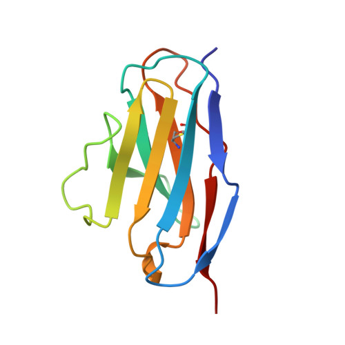
    1B0W, 1BZD, 1BZE, 1TSH, 2TRH, 2TRY

  • PubMed Abstract: 

    The most common form of hereditary systemic amyloidosis is familial amyloidotic polyneuropathy associated with single amino acid changes in the plasma protein transthyretin. So far, high resolution structures of only three amyloidogenic variants (Met30, Ser84, Ile122) and one non-amyloidogenic variant (Thr109) have been reported complemented by X-ray fiber diffraction studies and image reconstruction from electron micrographs of amyloid fibrils. To investigate the role of structural factors in this disease, we extended our studies to other transthyretin variants. We report crystallization and structural investigations of three amyloidogenic (Arg10, Ala60, Tyr77) and two non-amyloidogenic variants (Ser6, Met119). The similarity of these structures to normal transthyretin does not give direct clues to the fibril forming process. Since transthyretin amyloid fibrils contain a major fragment starting at position 49, besides the intact molecule, we calculated the solvent accessibility of residue 48. Indeed, all amyloidogenic variants show an increased main chain solvent exposure when compared to normal transthyretin and non-amyloidogenic variants, which can be postulated to result in increased susceptibility to proteolysis. After limited proteolysis, dimers are incapable of reassociation to native tetramers. We present a model for amyloid fibril formation based on formation of fibrils from N-terminal truncated dimers as building blocks.


  • Organizational Affiliation
    • Department of Medical and Molecular Genetics, Indiana University School of Medicine, Indianapolis 46202, USA.

Macromolecule Content 

  • Total Structure Weight: 35.84 kDa 
  • Atom Count: 2,624 
  • Modeled Residue Count: 324 
  • Deposited Residue Count: 324 
  • Unique protein chains: 1

Macromolecules

Find similar proteins by:|  3D Structure
Entity ID: 1
MoleculeChains  Sequence LengthOrganismDetailsImage
BENCE-JONES KAPPA I PROTEIN BRE
A, B, C
108Homo sapiensMutation(s): 0 
UniProt & NIH Common Fund Data Resources
Find proteins for P01594 (Homo sapiens)
Explore P01594 
Go to UniProtKB:  P01594
GTEx:  ENSG00000242076 
Entity Groups
Sequence Clusters30% Identity50% Identity70% Identity90% Identity95% Identity100% Identity
UniProt GroupP01594
Sequence Annotations
Expand
Reference Sequence

Experimental Data & Validation

Experimental Data

  • Method: X-RAY DIFFRACTION
  • Resolution: 1.80 Å
  • R-Value Free:  0.299 (Depositor) 
  • R-Value Work:  0.226 (Depositor), 0.229 (DCC) 
Space Group: C 2 2 21
Unit Cell:
Length ( Å )Angle ( ˚ )
a = 82.04α = 90
b = 142.11β = 90
c = 77.86γ = 90
Software Package:
Software NamePurpose
bioteXdata collection
bioteXdata reduction
AMoREphasing
X-PLORrefinement
bioteXdata scaling

Structure Validation

View Full Validation Report


Entry History 

Deposition Data

Revision History  (Full details and data files)

  • Version 1.0: 1998-11-16
    Type: Initial release
  • Version 1.1: 2008-04-26
    Changes: Version format compliance
  • Version 1.2: 2011-07-13
    Changes: Version format compliance
  • Version 1.3: 2017-10-04
    Changes: Refinement description
  • Version 1.4: 2018-05-30
    Changes: Advisory, Author supporting evidence, Data collection, Derived calculations
  • Version 1.5: 2023-08-09
    Changes: Data collection, Database references, Refinement description
  • Version 1.6: 2024-10-16
    Changes: Structure summary