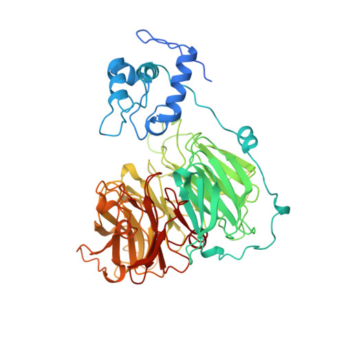
1AOF

CYTOCHROME CD1 NITRITE REDUCTASE, REDUCED FORM


Experimental Data Snapshot

  • Method: X-RAY DIFFRACTION
  • Resolution: 2.00 Å
  • R-Value Free: 0.201 
  • R-Value Work: 0.170 

wwPDB Validation   3D Report Full Report


Ligand Structure Quality Assessment 


This is version 1.3 of the entry. See complete history


Literature

Haem-ligand switching during catalysis in crystals of a nitrogen-cycle enzyme.

Williams, P.A.Fulop, V.Garman, E.F.Saunders, N.F.Ferguson, S.J.Hajdu, J.

(1997) Nature 389: 406-412

  • DOI: https://doi.org/10.1038/38775
  • Primary Citation of Related Structures:  
    1AOF, 1AOM, 1AOQ

  • PubMed Abstract: 

    Cytochrome cd1 nitrite reductase catalyses the conversion of nitrite to nitric oxide in the nitrogen cycle. The crystal structure of the oxidized enzyme shows that the d1 haem iron of the active site is ligated by His/Tyr side chains, and the c haem iron is ligated by a His/His ligand pair. Here we show that both haems undergo re-ligation during catalysis. Upon reduction, the tyrosine ligand of the d1 haem is released to allow substrate binding. Concomitantly, a refolding of the cytochrome c domain takes place, resulting in an unexpected change of the c haem iron coordination from His 17/His 69 to Met106/His69. This step is similar to the last steps in the folding of cytochrome c. The changes must affect the redox potential of the haems, and suggest a mechanism by which internal electron transfer is regulated. Structures of reaction intermediates show how nitric oxide is formed and expelled from the active-site iron, as well as how both haems return to their starting coordination. These results show how redox energy can be switched into conformational energy within a haem protein.


  • Organizational Affiliation

    Laboratory of Molecular Biophysics, University of Oxford, UK. pamela@scripps.edu


Macromolecules
Find similar proteins by:  (by identity cutoff)  |  3D Structure
Entity ID: 1
MoleculeChains Sequence LengthOrganismDetailsImage
NITRITE REDUCTASE
A, B
567Paracoccus pantotrophusMutation(s): 0 
UniProt
Find proteins for P72181 (Paracoccus pantotrophus)
Explore P72181 
Go to UniProtKB:  P72181
Entity Groups  
Sequence Clusters30% Identity50% Identity70% Identity90% Identity95% Identity100% Identity
UniProt GroupP72181
Sequence Annotations
Expand
  • Reference Sequence
Experimental Data & Validation

Experimental Data

  • Method: X-RAY DIFFRACTION
  • Resolution: 2.00 Å
  • R-Value Free: 0.201 
  • R-Value Work: 0.170 
  • Space Group: P 1 21 1
Unit Cell:
Length ( Å )Angle ( ˚ )
a = 107.4α = 90
b = 61.7β = 112.1
c = 101.2γ = 90
Software Package:
Software NamePurpose
X-PLORmodel building
X-PLORrefinement
DENZOdata reduction
SCALEPACKdata scaling
X-PLORphasing

Structure Validation

View Full Validation Report


Ligand Structure Quality Assessment 


Entry History 

Deposition Data

Revision History  (Full details and data files)

  • Version 1.0: 1997-10-15
    Type: Initial release
  • Version 1.1: 2008-03-24
    Changes: Version format compliance
  • Version 1.2: 2011-07-13
    Changes: Version format compliance
  • Version 1.3: 2024-06-05
    Changes: Data collection, Database references, Derived calculations, Other