1A0Q

29G11 COMPLEXED WITH PHENYL [1-(1-N-SUCCINYLAMINO)PENTYL] PHOSPHONATE


Experimental Data Snapshot

  • Method: X-RAY DIFFRACTION
  • Resolution: 2.30 Å
  • R-Value Free: 0.275 
  • R-Value Work: 0.203 
  • R-Value Observed: 0.203 

wwPDB Validation   3D Report Full Report


This is version 1.3 of the entry. See complete history


Literature

A comparison of the crystallographic structures of two catalytic antibodies with esterase activity.

Buchbinder, J.L.Stephenson, R.C.Scanlan, T.S.Fletterick, R.J.

(1998) J Mol Biol 282: 1033-1041

  • DOI: https://doi.org/10.1006/jmbi.1998.2025
  • Primary Citation of Related Structures:  
    1A0Q

  • PubMed Abstract: 

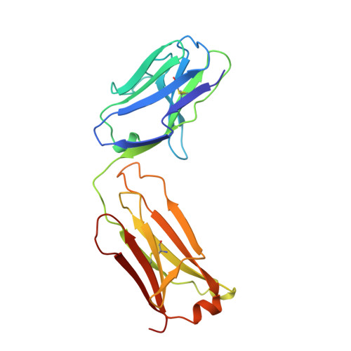
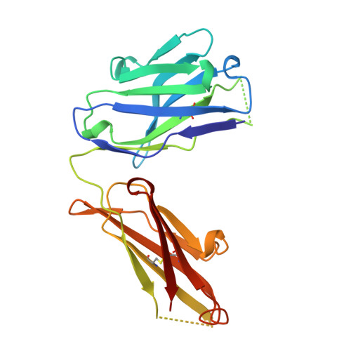
    The crystallographic structure of the Fab fragment of the catalytic antibody, 29G11, complexed with an (S)-norleucine phenyl phosphonate transition state analog was determined at 2.2 A resolution. The antibody catalyzes the hydrolysis of norleucine phenyl ester with (S)-enantioselectivity. The shape and charge complementarity of the binding pocket for the hapten account for the preferential binding of the (S)-enantiomer of the substrate. The structure is compared to that of the more catalytically efficient antibody, 17E8, induced by the same hapten transition state analog. 29G11 has different residues from 17E8 at eight positions in the heavy chain, including four substitutions in the hapten-binding pocket: A33V, S95G, S99R and Y100AN, and four substitutions at positions remote from the catalytic site, I28T, R40K, V65G and F91L. The two antibodies show large differences in the orientations of their variable and constant domains, reflected by a 32 degrees difference in their elbow angles. The VL and VH domains in the two antibodies differ by a rotation of 8.8 degrees. The hapten binds in similar orientations and locations in 29G11 and 17E8, which appear to have catalytic groups in common, though the changes in the association of the variable domains affect the precise positioning of residues in the hapten-binding pocket.


  • Organizational Affiliation

    Department of Biochemistry and Biophysics, University of California, San Francisco, San Francisco, CA, 94143-0448, USA.


Macromolecules
Find similar proteins by:  (by identity cutoff)  |  3D Structure
Entity ID: 1
MoleculeChains Sequence LengthOrganismDetailsImage
29G11 FAB (LIGHT CHAIN)A [auth L]212Mus musculusMutation(s): 0 
UniProt
Find proteins for P01837 (Mus musculus)
Explore P01837 
Go to UniProtKB:  P01837
Entity Groups  
Sequence Clusters30% Identity50% Identity70% Identity90% Identity95% Identity100% Identity
UniProt GroupP01837
Sequence Annotations
Expand
  • Reference Sequence
Find similar proteins by:  (by identity cutoff)  |  3D Structure
Entity ID: 2
MoleculeChains Sequence LengthOrganismDetailsImage
29G11 FAB (HEAVY CHAIN)B [auth H]217Mus musculusMutation(s): 0 
UniProt
Find proteins for P01869 (Mus musculus)
Explore P01869 
Go to UniProtKB:  P01869
Entity Groups  
Sequence Clusters30% Identity50% Identity70% Identity90% Identity95% Identity100% Identity
UniProt GroupP01869
Sequence Annotations
Expand
  • Reference Sequence
Small Molecules
Ligands 2 Unique
IDChains Name / Formula / InChI Key2D Diagram3D Interactions
HEP
Query on HEP

Download Ideal Coordinates CCD File 
F [auth H]PHENYL[1-(N-SUCCINYLAMINO)PENTYL]PHOSPHONATE
C15 H22 N O6 P
FJQWWGCHPFSERW-CQSZACIVSA-N
ZN
Query on ZN

Download Ideal Coordinates CCD File 
C [auth L],
D [auth H],
E [auth H]
ZINC ION
Zn
PTFCDOFLOPIGGS-UHFFFAOYSA-N
Binding Affinity Annotations 
IDSourceBinding Affinity
HEP PDBBind:  1A0Q Ki: 27 (nM) from 1 assay(s)
Binding MOAD:  1A0Q Ki: 27 (nM) from 1 assay(s)
Experimental Data & Validation

Experimental Data

  • Method: X-RAY DIFFRACTION
  • Resolution: 2.30 Å
  • R-Value Free: 0.275 
  • R-Value Work: 0.203 
  • R-Value Observed: 0.203 
  • Space Group: P 21 21 21
Unit Cell:
Length ( Å )Angle ( ˚ )
a = 36.78α = 90
b = 82.61β = 90
c = 132.34γ = 90
Software Package:
Software NamePurpose
DENZOdata reduction
SCALEPACKdata scaling
X-PLORmodel building
X-PLORrefinement
X-PLORphasing

Structure Validation

View Full Validation Report


Entry History 

Deposition Data

Revision History  (Full details and data files)

  • Version 1.0: 1999-03-02
    Type: Initial release
  • Version 1.1: 2008-03-24
    Changes: Version format compliance
  • Version 1.2: 2011-07-13
    Changes: Version format compliance
  • Version 1.3: 2023-08-02
    Changes: Database references, Derived calculations, Refinement description