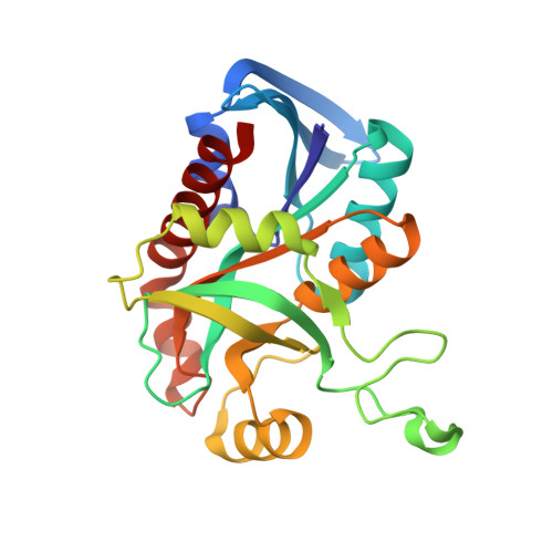
Structure and inhibition of a quorum sensing target from Streptococcus pneumoniae.
Singh, V., Shi, W., Almo, S.C., Evans, G.B., Furneaux, R.H., Tyler, P.C., Painter, G.F., Lenz, D.H., Mee, S., Zheng, R., Schramm, V.L.(2006) Biochemistry 45: 12929-12941
- PubMed: 17059210 Search on PubMedSearch on PubMed Central
- DOI: https://doi.org/10.1021/bi061184i
- Primary Citation Related Structures:
1ZOS - PubMed Abstract:
Streptococcus pneumoniae 5'-methylthioadenosine/S-adenosylhomocysteine hydrolase (MTAN) catalyzes the hydrolytic deadenylation of its substrates to form adenine and 5-methylthioribose or S-ribosylhomocysteine (SRH). MTAN is not found in mammals but is involved in bacterial quorum sensing. MTAN gene disruption affects the growth and pathogenicity of bacteria, making it a target for antibiotic design. Kinetic isotope effects and computational studies have established a dissociative S(N)1 transition state for Escherichia coli MTAN, and transition state analogues resembling the transition state are powerful inhibitors of the enzyme [Singh, V., Lee, J. L., Núñez, S., Howell, P. L., and Schramm, V. L. (2005) Biochemistry 44, 11647-11659]. The sequence of MTAN from S. pneumoniae is 40% identical to that of E. coli MTAN, but S. pneumoniae MTAN exhibits remarkably distinct kinetic and inhibitory properties. 5'-Methylthio-Immucillin-A (MT-ImmA) is a transition state analogue resembling an early S(N)1 transition state. It is a weak inhibitor of S. pneumoniae MTAN with a K(i) of 1.0 microM. The X-ray structure of S. pneumoniae MTAN with MT-ImmA indicates a dimer with the methylthio group in a flexible hydrophobic pocket. Replacing the methyl group with phenyl (PhT-ImmA), tolyl (p-TolT-ImmA), or ethyl (EtT-ImmA) groups increases the affinity to give K(i) values of 335, 60, and 40 nM, respectively. DADMe-Immucillins are geometric and electrostatic mimics of a fully dissociated transition state and bind more tightly than Immucillins. MT-DADMe-Immucillin-A inhibits with a K(i) value of 24 nM, and replacing the 5'-methyl group with p-Cl-phenyl (p-Cl-PhT-DADMe-ImmA) gave a K(i) value of 0.36 nM. The inhibitory potential of DADMe-Immucillins relative to the Immucillins supports a fully dissociated transition state structure for S. pneumoniae MTAN. Comparison of active site contacts in the X-ray crystal structures of E. coli and S. pneumoniae MTAN with MT-ImmA would predict equal binding, yet most analogues bind 10(3)-10(4)-fold more tightly to the E. coli enzyme. Catalytic site efficiency is primarily responsible for this difference since k(cat)/K(m) for S. pneumoniae MTAN is decreased 845-fold relative to that of E. coli MTAN.
- Department of Biochemistry, Albert Einstein College of Medicine, Bronx, New York 10461, USA.
Organizational Affiliation: