Conformation and mode of membrane interaction in cyclotides. Spatial structure of kalata B1 bound to a dodecylphosphocholine micelle.
Shenkarev, Z.O., Nadezhdin, K.D., Sobol, V.A., Sobol, A.G., Skjeldal, L., Arseniev, A.S.(2006) FEBS J 273: 2658-2672
- PubMed: 16817894 Search on PubMed
- DOI: https://doi.org/10.1111/j.1742-4658.2006.05282.x
- Primary Citation Related Structures:
1ZNU - PubMed Abstract:
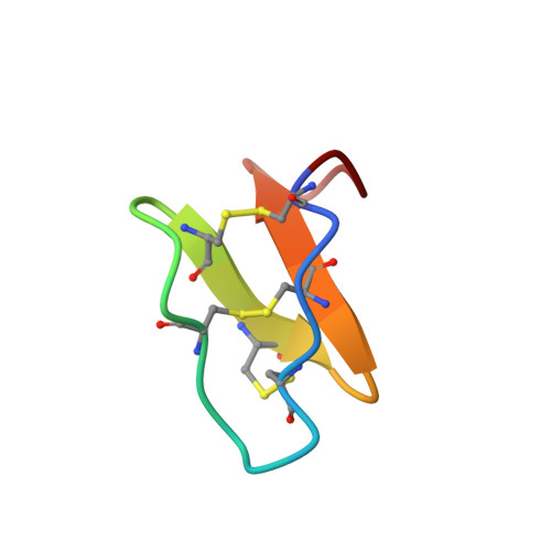
Cyclotides are a family of bioactive plant peptides that are characterized by a circular protein backbone and three conserved tightly packed disulfide bonds. The antimicrobial and hemolytic properties of cyclotides, along with the relative hydrophobicity of the peptides, point to the biological membrane as a target for cyclotides. To assess the membrane-induced conformation and orientation of cyclotides, the interaction of the Möbius cyclotide, kalata B1, from the African perennial plant Oldenlandia affinis, with dodecylphosphocholine micelles was studied using NMR spectroscopy. Under conditions where the cyclotide formed a well-defined complex with micelles, the spatial structure of kalata B1 was calculated from NOE and J couplings data, and the model for the peptide-micelle complex was built using 5- and 16-doxylstearate relaxation probes. The binding of divalent cations to the peptide-micelle complex was quantified by Mn2+ titration. The results show that the peptide binds to the micelle surface, with relatively high affinity, via two hydrophobic loops (loop 5, Trp19-Val21; and loop6, Leu27-Val29). The charged residues (Glu3 and Arg24), along with the cation-binding site (near Glu3) are segregated on the other side of the molecule and in contact with polar head groups of detergent. The spatial structure of kalata B1 is only slightly changed during incorporation into micelles and represents a distorted triple-stranded beta-sheet cross-linked by a cystine knot. Detailed structural analysis and comparison with other knottins revealed structural conservation of the two-disulfide motif in cyclic and acyclic peptides. The results thus obtained provide the first model for interaction of cyclotides with membranes and permit consideration of the cyclotides as membrane-active cationic antimicrobial peptides.
- Shemyakin-Ovchinnikov Institute of Bioorganic Chemistry, Russian Academy of Sciences, Moscow, Russia.
Organizational Affiliation: