1ZK9

NF-kB RelB forms an intertwined homodimer


Experimental Data Snapshot

  • Method: X-RAY DIFFRACTION
  • Resolution: 2.18 Å
  • R-Value Free: 0.218 
  • R-Value Work: 0.195 
  • R-Value Observed: 0.195 

wwPDB Validation   3D Report Full Report


This is version 1.4 of the entry. See complete history


Literature

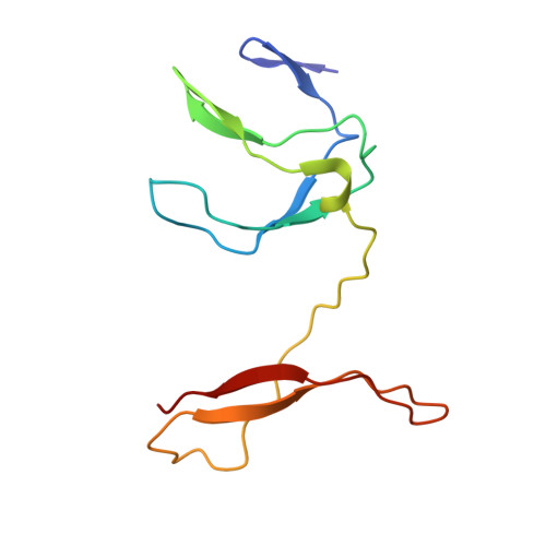
NF-kappaB RelB Forms an Intertwined Homodimer.

Huang, D.B.Vu, D.Ghosh, G.

(2005) Structure 13: 1365-1373

  • DOI: https://doi.org/10.1016/j.str.2005.06.018
  • Primary Citation of Related Structures:  
    1ZK9, 1ZKA

  • PubMed Abstract: 

    The X-ray structure of the RelB dimerization domain (DD) reveals that the RelBDD assumes an unexpected intertwined fold topology atypical of other NF-kappaB dimers. All typical NF-kappaB dimers are formed by the association of two independently folded immunoglobulin (Ig) domains. In RelBDD, two polypeptides reconstruct both Ig domains in the dimer with an extra beta sheet connecting the two domains. Residues most critical to NF-kappaB dimer formation are invariant in RelB, and Y300 plays a positive role in RelBDD dimer formation. The presence of RelB-specific nonpolar residues at the surface removes several intradomain surface hydrogen bonds that may render the domain fold unstable. Intertwining may stabilize the RelBDD homodimer by forming the extra beta sheet. We show that, as in the crystal, RelB forms an intertwined homodimer in solution. We suggest that the transiently stable RelB homodimer might prevent its rapid degradation, allowing for heterodimer formation with p50 and p52.


  • Organizational Affiliation

    Department of Chemistry and Biochemistry, University of California, San Diego, 9500 Gilman Drive, La Jolla, CA 92093, USA.


Macromolecules
Find similar proteins by:  (by identity cutoff)  |  3D Structure
Entity ID: 1
MoleculeChains Sequence LengthOrganismDetailsImage
Transcription factor RelB110Mus musculusMutation(s): 0 
Gene Names: Relb
UniProt
Find proteins for Q04863 (Mus musculus)
Explore Q04863 
Go to UniProtKB:  Q04863
Entity Groups  
Sequence Clusters30% Identity50% Identity70% Identity90% Identity95% Identity100% Identity
UniProt GroupQ04863
Sequence Annotations
Expand
  • Reference Sequence
Experimental Data & Validation

Experimental Data

  • Method: X-RAY DIFFRACTION
  • Resolution: 2.18 Å
  • R-Value Free: 0.218 
  • R-Value Work: 0.195 
  • R-Value Observed: 0.195 
  • Space Group: P 31 2 1
Unit Cell:
Length ( Å )Angle ( ˚ )
a = 69.2α = 90
b = 69.2β = 90
c = 64.74γ = 120
Software Package:
Software NamePurpose
CNSrefinement
DENZOdata reduction
SCALEPACKdata scaling
AMoREphasing

Structure Validation

View Full Validation Report


Entry History 

Deposition Data

Revision History  (Full details and data files)

  • Version 1.0: 2005-05-10
    Type: Initial release
  • Version 1.1: 2008-04-30
    Changes: Version format compliance
  • Version 1.2: 2011-07-13
    Changes: Derived calculations, Version format compliance
  • Version 1.3: 2018-04-04
    Changes: Data collection
  • Version 1.4: 2023-08-23
    Changes: Data collection, Database references, Refinement description