1ZJA

Crystal structure of the trehalulose synthase MutB from Pseudomonas mesoacidophila MX-45 (triclinic form)


Experimental Data Snapshot

  • Method: X-RAY DIFFRACTION
  • Resolution: 1.60 Å
  • R-Value Free: 0.219 
  • R-Value Work: 0.184 
  • R-Value Observed: 0.184 

wwPDB Validation   3D Report Full Report


This is version 1.2 of the entry. See complete history


Literature

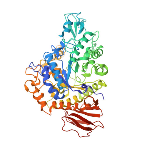
Trehalulose synthase native and carbohydrate complexed structures provide insights into sucrose isomerization.

Ravaud, S.Robert, X.Watzlawick, H.Haser, R.Mattes, R.Aghajari, N.

(2007) J Biol Chem 282: 28126-28136

  • DOI: 10.1074/jbc.M704515200
  • Primary Citation of Related Structures:  
    1ZJA, 2PWD, 2PWE, 2PWF, 2PWG, 2PWH

  • PubMed Abstract: 
  • Various diseases related to the overconsumption of sugar make a growing need for sugar substitutes. Because sucrose is an inexpensive and readily available d-glucose donor, the industrial potential for enzymatic synthesis of the sucrose isomers trehalulose and/or isomaltulose from sucrose is large ...

    Various diseases related to the overconsumption of sugar make a growing need for sugar substitutes. Because sucrose is an inexpensive and readily available d-glucose donor, the industrial potential for enzymatic synthesis of the sucrose isomers trehalulose and/or isomaltulose from sucrose is large. The product specificity of sucrose isomerases that catalyze this reaction depends essentially on the possibility for tautomerization of sucrose, which is required for trehalulose formation. For optimal use of the enzyme, targeting controlled synthesis of these functional isomers, it is necessary to minimize the side reactions. This requires an extensive analysis of substrate binding modes and of the specificity-determining sites in the structure. The 1.6-2.2-A resolution three-dimensional structures of native and mutant complexes of a trehalulose synthase from Pseudomonas mesoacidophila MX-45 mimic successive states of the enzyme reaction. Combined with mutagenesis studies they give for the first time thorough insights into substrate recognition and processing and reaction specificities of these enzymes. Among the important outcomes of this study is the revelation of an aromatic clamp defined by Phe(256) and Phe(280) playing an essential role in substrate recognition and in controlling the reaction specificity, which is further supported by mutagenesis studies. Furthermore, this study highlights essential residues for binding the glucosyl and fructosyl moieties. The introduction of subtle changes informed by comparative three-dimensional structural data observed within our study can lead to fundamental modifications in the mode of action of sucrose isomerases and hence provide a template for industrial catalysts.


    Related Citations: 
    • Expression, purification, crystallization and preliminary X-ray crystallographic studies of the trehalulose synthase MutB from Pseudomonas mesoacidophila MX-45.
      Ravaud, S., Watzlawick, H., Haser, R., Mattes, R., Aghajari, N.
      (2005) Acta Crystallogr Sect F Struct Biol Cryst Commun 61: 100

    Organizational Affiliation

    Laboratoire de BioCristallographie, Institut de Biologie et Chimie des Protéines, CNRS et Université de Lyon, UMR 5086, IFR 128 BioSciences Gerland-Lyon Sud, F-69367 Lyon Cedex 07, France.


Macromolecules
Find similar proteins by:  (by identity cutoff)  |  3D Structure
Entity ID: 1
MoleculeChainsSequence LengthOrganismDetailsImage
Trehalulose synthaseA, B557Burkholderia ubonensis subsp. mesacidophilaMutation(s): 0 
Gene Names: mutB
EC: 5.4.99.11
UniProt
Find proteins for Q2PS28 (Burkholderia ubonensis subsp. mesacidophila)
Explore Q2PS28 
Go to UniProtKB:  Q2PS28
Entity Groups  
Sequence Clusters30% Identity50% Identity70% Identity90% Identity95% Identity100% Identity
UniProt GroupQ2PS28
Protein Feature View
Expand
  • Reference Sequence
Small Molecules
Ligands 2 Unique
IDChainsName / Formula / InChI Key2D Diagram3D Interactions
TRS
Query on TRS

Download Ideal Coordinates CCD File 
D [auth A],
F [auth B]
2-AMINO-2-HYDROXYMETHYL-PROPANE-1,3-DIOL
C4 H12 N O3
LENZDBCJOHFCAS-UHFFFAOYSA-O
 Ligand Interaction
CA
Query on CA

Download Ideal Coordinates CCD File 
C [auth A],
E [auth B]
CALCIUM ION
Ca
BHPQYMZQTOCNFJ-UHFFFAOYSA-N
 Ligand Interaction
Experimental Data & Validation

Experimental Data

  • Method: X-RAY DIFFRACTION
  • Resolution: 1.60 Å
  • R-Value Free: 0.219 
  • R-Value Work: 0.184 
  • R-Value Observed: 0.184 
  • Space Group: P 1
Unit Cell:
Length ( Å )Angle ( ˚ )
a = 63.67α = 67.36
b = 71.96β = 73.13
c = 82.24γ = 70.82
Software Package:
Software NamePurpose
CNSrefinement
MOSFLMdata reduction
SCALAdata scaling
AMoREphasing

Structure Validation

View Full Validation Report


Entry History 

Deposition Data

Revision History  (Full details and data files)

  • Version 1.0: 2006-10-17
    Type: Initial release
  • Version 1.1: 2007-10-16
    Changes: Version format compliance
  • Version 1.2: 2011-07-13
    Changes: Version format compliance