1ZA0 | pdb_00001za0

X-ray structure of putative acyl-ACP desaturase DesA2 from Mycobacterium tuberculosis H37Rv


Experimental Data Snapshot

  • Method: X-RAY DIFFRACTION
  • Resolution: 2.00 Å
  • R-Value Free: 
    0.237 (Depositor), 0.239 (DCC) 
  • R-Value Work: 
    0.206 (Depositor), 0.213 (DCC) 
  • R-Value Observed: 
    0.208 (Depositor) 

wwPDB Validation 3D Report Full Report

Validation slider image for 1ZA0

This is version 1.3 of the entry. See complete history

Literature

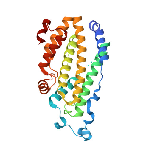
X-ray structure of putative acyl-ACP desaturase DesA2 from Mycobacterium tuberculosis H37Rv.

Dyer, D.H.Lyle, K.S.Rayment, I.Fox, B.G.

(2005) Protein Sci 14: 1508-1517

  • DOI: https://doi.org/10.1110/ps.041288005
  • Primary Citation Related Structures: 
    1ZA0

  • PubMed Abstract: 

    Genome sequencing showed that two proteins in Mycobacterium tuberculosis H37Rv contain the metal binding motif (D/E)X(2)HX(approximately 100)(D/E)X(2)H characteristic of the soluble diiron enzyme superfamily. These putative acyl-ACP desaturase genes desA1 and desA2 were cloned from genomic DNA and expressed in Escherichia coli BL21(DE3). DesA1 was found to be insoluble, but in contrast, DesA2 was a soluble protein amenable to biophysical characterization. Here, we report the 2.0 A resolution X-ray structure of DesA2 determined by multiple anomalous dispersion (MAD) phasing from a Se-met derivative and refinement against diffraction data obtained on the native protein. The X-ray structure shows that DesA2 is a homodimeric protein with a four-helix bundle core flanked by five additional helices that overlay with 192 structurally equivalent amino acids in the structure of stearoyl-ACP Delta9 desaturase from castor plant with an rms difference 1.42 A. In the DesA2 crystals, one metal (likely Mn from the crystallization buffer) was bound in high occupancy at the B-site of the conserved metal binding motif, while the A-site was not occupied by a metal ion. Instead, the amino group of Lys-76 occupied this position. The relationships between DesA2 and known diiron enzymes are discussed.


  • Organizational Affiliation
    • Department of Biochemistry, 433 Babcock Dr., University of Wisconsin, Madison, Madison, WI 53706, USA.

Macromolecule Content 

  • Total Structure Weight: 31.51 kDa 
  • Atom Count: 2,029 
  • Modeled Residue Count: 242 
  • Deposited Residue Count: 275 
  • Unique protein chains: 1

Macromolecules

Find similar proteins by:|  3D Structure
Entity ID: 1
MoleculeChains  Sequence LengthOrganismDetailsImage
POSSIBLE ACYL-[ACYL-CARRIER PROTEIN] DESATURASE DESA2 (ACYL-[ACP] DESATURASE) (STEAROYL-ACP DESATURASE)275Mycobacterium tuberculosis H37RvMutation(s): 0 
Gene Names: DesA2
EC: 1.14.19.2 (PDB Primary Data), 1.14.19 (UniProt)
UniProt
Find proteins for P9WNZ5 (Mycobacterium tuberculosis (strain ATCC 25618 / H37Rv))
Explore P9WNZ5 
Go to UniProtKB:  P9WNZ5
Entity Groups
Sequence Clusters30% Identity50% Identity70% Identity90% Identity95% Identity100% Identity
UniProt GroupP9WNZ5
Sequence Annotations
Expand
Reference Sequence

Experimental Data & Validation

Experimental Data

  • Method: X-RAY DIFFRACTION
  • Resolution: 2.00 Å
  • R-Value Free:  0.237 (Depositor), 0.239 (DCC) 
  • R-Value Work:  0.206 (Depositor), 0.213 (DCC) 
  • R-Value Observed: 0.208 (Depositor) 
Space Group: P 65 2 2
Unit Cell:
Length ( Å )Angle ( ˚ )
a = 66.1α = 90
b = 66.1β = 90
c = 292γ = 120
Software Package:
Software NamePurpose
REFMACrefinement
HKL-2000data reduction
SCALEPACKdata scaling
SOLVEphasing

Structure Validation

View Full Validation Report


Entry History 

Deposition Data

Revision History  (Full details and data files)

  • Version 1.0: 2005-06-07
    Type: Initial release
  • Version 1.1: 2008-04-30
    Changes: Version format compliance
  • Version 1.2: 2011-07-13
    Changes: Derived calculations, Source and taxonomy, Version format compliance
  • Version 1.3: 2024-02-14
    Changes: Data collection, Database references, Derived calculations