1Z9N

X-Ray structure of a Cu-Zn superoxide dismutase from Haemophilus ducreyi with haem bound at the dimer interface


Experimental Data Snapshot

  • Method: X-RAY DIFFRACTION
  • Resolution: 1.50 Å
  • R-Value Free: 0.193 
  • R-Value Work: 0.157 
  • R-Value Observed: 0.158 

wwPDB Validation   3D Report Full Report


Ligand Structure Quality Assessment 


This is version 1.4 of the entry. See complete history


Literature

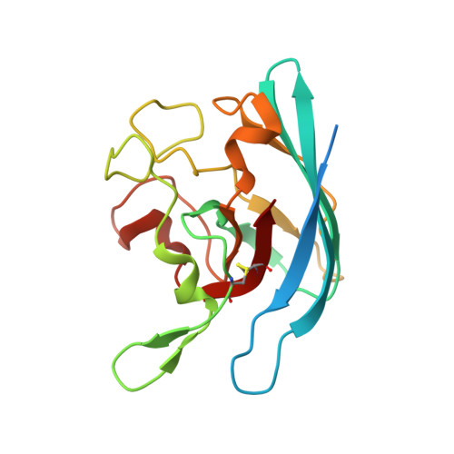
Structural basis of heme binding in the Cu,Zn superoxide dismutase from Haemophilus ducreyi.

Toro, I.Petrutz, C.Pacello, F.D'Orazio, M.Battistoni, A.Djinovic-Carugo, K.

(2009) J Mol Biol 386: 406-418

  • DOI: https://doi.org/10.1016/j.jmb.2008.12.004
  • Primary Citation of Related Structures:  
    1Z9N, 1Z9P

  • PubMed Abstract: 

    The Cu,Zn superoxide dismutase from Haemophilus ducreyi is characterized by the unique ability to bind heme at its dimer interface. Here we report the high-resolution crystal structures of this protein in the heme-loaded (holo) and heme-free (apo) forms. Heme is asymmetrically bound between the two enzyme subunits, where heme iron is coordinated by two histidine residues, His64 and His 124, provided by the two subunits. Moreover, the binding of heme to the protein is ensured by stabilizing contacts between the prosthetic group and a limited number of other residues, most of which are not present in other bacterial enzyme variants. We show that the introduction of only three mutations at the dimer interface of the enzyme from Haemophilus parainfluenzae, a closely related bacterial species, is sufficient to induce heme-binding ability by this enzyme variant. Heme binding does not alter protein activity. Moreover, the binding of the prosthetic group does not induce any significant structural perturbation at the subunit level and requires only limited local structural rearrangements that widen the cleft at the dimer interface and cause a limited shift in the relative orientation between the subunits. The presence of a preformed heme-binding pocket and the significant solvent exposure of the cofactor to the solvent are compatible with the suggested protective role of the enzyme against heme toxicity or with its involvement in heme trafficking in the periplasmic space.


  • Organizational Affiliation

    Structural and Computational Biology Programme, EMBL-Heidelberg, Germany.


Macromolecules
Find similar proteins by:  (by identity cutoff)  |  3D Structure
Entity ID: 1
MoleculeChains Sequence LengthOrganismDetailsImage
Superoxide dismutase [Cu-Zn]
A, B, C, D
177[Haemophilus] ducreyiMutation(s): 0 
Gene Names: sodC
EC: 1.15.1.1
UniProt
Find proteins for Q59452 (Haemophilus ducreyi (strain 35000HP / ATCC 700724))
Explore Q59452 
Go to UniProtKB:  Q59452
Entity Groups  
Sequence Clusters30% Identity50% Identity70% Identity90% Identity95% Identity100% Identity
UniProt GroupQ59452
Sequence Annotations
Expand
  • Reference Sequence
Small Molecules
Ligands 3 Unique
IDChains Name / Formula / InChI Key2D Diagram3D Interactions
HEM
Query on HEM

Download Ideal Coordinates CCD File 
G [auth A],
L [auth C]
PROTOPORPHYRIN IX CONTAINING FE
C34 H32 Fe N4 O4
KABFMIBPWCXCRK-RGGAHWMASA-L
ZN
Query on ZN

Download Ideal Coordinates CCD File 
F [auth A],
I [auth B],
K [auth C],
N [auth D]
ZINC ION
Zn
PTFCDOFLOPIGGS-UHFFFAOYSA-N
CU
Query on CU

Download Ideal Coordinates CCD File 
E [auth A],
H [auth B],
J [auth C],
M [auth D]
COPPER (II) ION
Cu
JPVYNHNXODAKFH-UHFFFAOYSA-N
Experimental Data & Validation

Experimental Data

  • Method: X-RAY DIFFRACTION
  • Resolution: 1.50 Å
  • R-Value Free: 0.193 
  • R-Value Work: 0.157 
  • R-Value Observed: 0.158 
  • Space Group: P 1
Unit Cell:
Length ( Å )Angle ( ˚ )
a = 37.32α = 66.59
b = 65.9β = 89.87
c = 69.54γ = 76.03
Software Package:
Software NamePurpose
XDSdata reduction
AMoREphasing
REFMACrefinement
XDSdata scaling

Structure Validation

View Full Validation Report


Ligand Structure Quality Assessment 


Entry History 

Deposition Data

Revision History  (Full details and data files)

  • Version 1.0: 2006-09-19
    Type: Initial release
  • Version 1.1: 2008-04-30
    Changes: Version format compliance
  • Version 1.2: 2011-07-13
    Changes: Version format compliance
  • Version 1.3: 2011-10-26
    Changes: Database references
  • Version 1.4: 2023-08-23
    Changes: Data collection, Database references, Derived calculations, Refinement description